V体育ios版 - Skip to main page content
U.S. flag

An official website of the United States government

Dot gov

The . gov means it’s official. Federal government websites often end in . gov or VSports app下载. mil. Before sharing sensitive information, make sure you’re on a federal government site. .

Https

The site is secure V体育官网. The https:// ensures that you are connecting to the official website and that any information you provide is encrypted and transmitted securely. .

. 2011 Apr 15;286(15):13754-64.
doi: 10.1074/jbc.M110.194936. Epub 2011 Feb 24.

Fbxw7-dependent degradation of Notch is required for control of "stemness" and neuronal-glial differentiation in neural stem cells

Affiliations

Fbxw7-dependent degradation of Notch is required for control of "stemness" and neuronal-glial differentiation in neural stem cells

Akinobu Matsumoto et al. J Biol Chem. .

Abstract

Control of the growth and differentiation of neural stem cells is fundamental to brain development and is largely dependent on the Notch signaling pathway VSports手机版. The mechanism by which the activity of Notch is regulated during brain development has remained unclear, however. Fbxw7 (also known as Fbw7, SEL-10, hCdc4, or hAgo) is the F-box protein subunit of an Skp1-Cul1-F-box protein (SCF)-type ubiquitin ligase complex that plays a central role in the degradation of Notch family members. We now show that mice with brain-specific deletion of Fbxw7 (Nestin-Cre/Fbxw7(F/F) mice) die shortly after birth with morphological abnormalities of the brain and the absence of suckling behavior. The maintenance of neural stem cells was sustained in association with the accumulation of Notch1 and Notch3, as well as up-regulation of Notch target genes in the mutant mice. Astrogenesis was also enhanced in the mutant mice in vivo, and the differentiation of neural progenitor cells was skewed toward astrocytes rather than neurons in vitro, with the latter effect being reversed by treatment of the cells with a pharmacological inhibitor of the Notch signaling pathway. Our results thus implicate Fbxw7 as a key regulator of the maintenance and differentiation of neural stem cells in the brain. .


Figures (VSports最新版本)

FIGURE 1.
FIGURE 1.
Generation of conditional knockout mice lacking Fbxw7 specifically in the nervous system. A, PCR analysis of genomic DNA from the brain of mice of the indicated genotypes at P0.5. The positions of amplified fragments corresponding to wild-type (WT), floxed (Flox), and ΔE5 (exon 5-deleted) alleles are indicated. B, RT and real-time PCR analysis of Fbxw7 mRNA in the brain of Fbxw7+/F, Nestin-Cre/Fbxw7+/F, and Nestin-Cre/Fbxw7F/F mice at E14.5. Normalized data are expressed relative to the value for Fbxw7+/F mice and are mean ± S.D. from three independent experiments. C, gross appearance of newborns and the neonatal stomach of Nestin-Cre/Fbxw7F/F (Fbxw7 CKO) and control mice. The arrows indicate the position of the stomach. Scale bars = 5 mm. D, summary of suckling behavior of Nestin-Cre/Fbxw7+/F (Control) and Fbxw7 CKO mice at P0.5. E, H&E staining of lung tissue from Nestin-Cre/Fbxw7+/F (Control) and Fbxw7 CKO mice at P0.5. Scale bars = 100 μm.
FIGURE 2.
FIGURE 2.
Morphological abnormalities of the brain of Nestin-Cre/Fbxw7F/F mice. A and B, H&E staining of coronal sections (A) and sagittal sections (B) of the brain of Nestin-Cre/Fbxw7+/F (Control) and Nestin-Cre/Fbxw7F/F (Fbxw7 CKO) mice at E16.5 and E18.5 (A) or P0.5 (B). The arrows in (A) indicate a horizontal sulcus characteristic of the mutant mice, which is shown at higher magnification in the inset. The arrow and arrowhead in (B) indicate the pons and cerebellum, respectively. Scale bars = 500 μm (A) and 1 mm (B). C, H&E staining of coronal sections of the cortex of Nestin-Cre/Fbxw7+/F (Control) and Fbxw7 CKO mice at E18.5. CP, cortical plate; IZ, intermediate zone; VZ, ventricular zone; SVZ, subventricular zone. Scale bars = 100 μm.
FIGURE 3.
FIGURE 3.
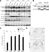
Notch accumulation in the Fbxw7-deficient brain. A, immunoblot (IB) analysis of Fbxw7 substrates and HSP70 (loading control) in brain extracts from Nestin-Cre/Fbxw7+/F (Control) and Nestin-Cre/Fbxw7F/F (Fbxw7 CKO) mice at E12.5 to P0.5. The amounts of the Notch intracellular domain (NICD) of Notch1 or Notch3 (both phosphorylated and nonphosphorylated forms) in the mutant brain relative to those in the control brain at each developmental stage were determined by densitometry and are shown below each lane. mTOR, mammalian target of rapamycin. B, protein extracts from the brain of Nestin-Cre/Fbxw7+/F (Control) and Fbxw7 CKO mice at E14.5 were incubated with or without calf intestinal alkaline phosphatase (CIAP) for 1 h before immunoblot analysis of Notch1. C, RT and real-time PCR analysis of Notch target gene expression in the brain of Nestin-Cre/Fbxw7+/F (Control) and Fbxw7 CKO mice at E14.5. Normalized data are expressed relative to the corresponding value for control mice and are mean ± S.D. from three independent experiments. *, p < 0.05; **, p < 0.01; N.S., not significant. D, in situ hybridization analysis of Hes5 mRNA in the brain of Nestin-Cre/Fbxw7+/F (Control) and Fbxw7 CKO mice at E14.5. Scale bars = 500 μm.
FIGURE 4.
FIGURE 4.
Impairment of neuronal differentiation and enhanced astroglial differentiation in the Fbxw7-deficient brain. A, immunofluorescence analysis of βIII-tubulin (green) and GFAP (red) in primary cultured cells from the cerebrum of Nestin-Cre/Fbxw7+/F (Control) and Nestin-Cre/Fbxw7F/F (Fbxw7 CKO) mice at P0.5. Nuclei were stained blue with Hoechst 33258. Scale bars = 40 μm. B, quantitative analysis of cells positive for βIII-tubulin, GFAP, or MBP in experiments similar to that in A. Data are mean ± S.D. from three independent experiments. **, p < 0.01; ***, p < 0.005; N.S., not significant. C, immunofluorescence analysis of BrdU (green) and Ki67 (red) in the brain of Nestin-Cre/Fbxw7+/F (Control) and Fbxw7 CKO mice at 30 h after labeling with a single pulse of BrdU at E13.25. More BrdU-positive cells had migrated into the Ki67-negative differentiated mantle zone (MZ) of control mice than into that of Fbxw7 CKO mice. Scale bars = 100 μm (left panels) and 50 μm (right panels). D, quantitative analysis of cells positive for BrdU in the mantle zone in experiments similar to that in C. Data are mean ± S.D. from three independent experiments. ***, p < 0.005. E, immunofluorescence analysis of GFAP in the posterior midbrain of Nestin-Cre/Fbxw7+/F (Control) and Fbxw7 CKO mice at P0.5. Scale bars = 100 μm.
FIGURE 5.
FIGURE 5.
Promotion of neural stem cell proliferation by loss of Fbxw7. A, immunofluorescence analysis of pHH3 (green) in the brain of Nestin-Cre/Fbxw7+/F (Control) and Nestin-Cre/Fbxw7F/F (Fbxw7 CKO) mice at E14.5 and E16.5. Nuclei were stained blue with Hoechst 33258. Scale bars = 50 μm. B, quantitative analysis of cells positive for pHH3 in the ventricular zone (VZ) or in the subventricular zone (SVZ) and intermediate zone (IZ) per image area in experiments similar to that in A. Data are mean ± S.D. from four independent experiments. ***, p < 0.005; N.S., not significant. C, immunofluorescence analysis of Tbr2 (green) in the brain of Nestin-Cre/Fbxw7+/F (Control) and Fbxw7 CKO mice at E14.5 and E16.5. Nuclei were stained blue with Hoechst 33258. Scale bars = 50 μm. D, quantitative analysis of cells positive for Tbr2 per image area in experiments similar to that in C. Data are mean ± S.D. from four independent experiments. ***, p < 0.005.
FIGURE 6.
FIGURE 6.
Increased efficiency of neurosphere formation associated with Notch1 accumulation resulting from the loss of Fbxw7. A, light microscopy of neurospheres (tertiary) derived from the telencephalon of E14.5 Nestin-Cre/Fbxw7+/F (Control) and Nestin-Cre/Fbxw7F/F (Fbxw7 CKO) mice. Scale bars = 200 μm. B, numbers of secondary and tertiary neurospheres formed by 4000 dissociated cells from Nestin-Cre/Fbxw7+/F (Control) and Fbxw7 CKO mice. Data are mean ± S.D. from four and three independent experiments for secondary and tertiary neurospheres, respectively. *, p < 0.05. C, immunoblot (IB) analysis of Notch1 and HSP90 (loading control) in neurospheres (tertiary, 7 days after replating) and brain extracts (E14.5) derived from Nestin-Cre/Fbxw7+/F (Control) and Fbxw7 CKO mice. The amounts of the Notch intracellular domain (NICD) of Notch1 (both phosphorylated and nonphosphorylated forms) for the mutant genotype relative to those for the control genotype were determined by densitometry and are shown below each lane. D, immunoblot analysis of other Fbxw7 substrates in neurospheres (tertiary) from Nestin-Cre/Fbxw7+/F (Control) and Fbxw7 CKO mice at 7 days after replating. E, RT and real-time PCR analysis of Notch target gene expression in tertiary neurospheres from Nestin-Cre/Fbxw7+/F (Control) and Fbxw7 CKO mice at 3 days after replating. Normalized data are expressed relative to the corresponding value for control mice and are mean ± S.D. from three independent experiments. *, p < 0.05; ***, p < 0.005.
FIGURE 7.
FIGURE 7.
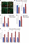
Attenuation of the abnormalities in the differentiation and maintenance of neural stem cells of Fbxw7-deficient mice by inhibition of Notch signaling. A, immunofluorescence analysis of βIII-tubulin (green) and GFAP (red) in primary cultured cells derived from the cerebrum of Nestin-Cre/Fbxw7+/F (Control) and Nestin-Cre/Fbxw7F/F (Fbxw7 CKO) mice at E18.5. The cells were cultured with 0.5 μm DAPT or 0.05% dimethyl sulfoxide (DMSO) vehicle for 7 days. Nuclei were stained blue with Hoechst 33258. Scale bars = 40 μm. B, quantitative analysis of cells positive for βIII-tubulin or GFAP in experiments similar to that in A. Data are mean ± S.D. from three independent experiments. *, p < 0.05; ***, p < 0.005; N.S., not significant. C, RT and real-time PCR analysis of Notch target gene expression in primary cultured cells from Nestin-Cre/Fbxw7+/F (Control) and Fbxw7 CKO mice. The cells were cultured with or without 0.5 μm DAPT for 2 days. Normalized data are expressed relative to the corresponding value for control cells treated with dimethyl sulfoxide and are mean ± S.D. from three independent experiments. ***, p < 0.005. D, number of tertiary neurospheres formed by 6000 dissociated cells derived from Nestin-Cre/Fbxw7+/F (Control) or Fbxw7 CKO mice and cultured with or without 0.5 μm DAPT. Data are mean ± S.D. from three independent experiments. *, p < 0.05; ***, p < 0.005. E, RT and real-time PCR analysis of Notch target gene expression in tertiary neurospheres derived from Nestin-Cre/Fbxw7+/F (Control) or Fbxw7 CKO mice. Dissociated cells were cultured with or without 0.5 μm DAPT for 3 days. Normalized data are expressed relative to the corresponding value for control cells treated with DMSO and are mean ± S.D. from three independent experiments. ***, p < 0.005; N.S., not significant.
FIGURE 8.
FIGURE 8.
Model for Fbxw7-mediated regulation of Notch signaling and neural development. Fbxw7 regulates the abundance of Notch by mediating its phosphorylation-dependent degradation. Loss of Fbxw7 results in the accumulation of Notch, an increase in the number of neural stem cells, and skewed differentiation of these cells into astrocytes, leading to aberrant brain development.

References

    1. Artavanis-Tsakonas S., Rand M. D., Lake R. J. (1999) Science 284, 770–776 - PubMed
    1. Justice N. J., Jan Y. N. (2002) Curr. Opin. Neurobiol. 12, 64–70 - PubMed
    1. Lai E. C. (2004) Development 131, 965–973 - PubMed
    1. Schuurmans C., Guillemot F. (2002) Curr. Opin. Neurobiol. 12, 26–34 - PubMed
    1. Furukawa T., Mukherjee S., Bao Z. Z., Morrow E. M., Cepko C. L. (2000) Neuron 26, 383–394 - PubMed (VSports)

Publication types

MeSH terms (VSports注册入口)