VSports app下载 - Skip to main page content
U.S. flag

An official website of the United States government

Dot gov

The . gov means it’s official VSports app下载. Federal government websites often end in . gov or . mil. Before sharing sensitive information, make sure you’re on a federal government site. .

Https

The site is secure. The https:// ensures that you are connecting to the official website and that any information you provide is encrypted and transmitted securely V体育官网. .

. 2011 Feb 14;208(2):295-312.
doi: 10.1084/jem.20100830. Epub 2011 Jan 31.

FBXW7 influences murine intestinal homeostasis and cancer, targeting Notch, Jun, and DEK for degradation

Affiliations

"V体育官网" FBXW7 influences murine intestinal homeostasis and cancer, targeting Notch, Jun, and DEK for degradation

Roya Babaei-Jadidi et al. J Exp Med. .

Abstract

The Fbxw7 (F-box/WD repeat-containing protein 7; also called CDC4, Sel10, Ago, and Fbw7) component of the SCF (Skp1/Cullin/F-box protein) E3 ubiquitin ligase complex acts as a tumor suppressor in several tissues and targets multiple transcriptional activators and protooncogenes for ubiquitin-mediated degradation. To understand Fbxw7 function in the murine intestine, in this study, we specifically deleted Fbxw7 in the murine gut using Villin-Cre (Fbxw7(ΔG)). In wild-type mice, loss of Fbxw7 in the gut altered homeostasis of the intestinal epithelium, resulted in elevated Notch and c-Jun expression, and induced development of adenomas at 9-10 mo of age. In the context of APC (adenomatous polyposis coli) deficiency (Apc(Min/+) mice), loss of Fbxw7 accelerated intestinal tumorigenesis and death and promoted accumulation of β-catenin in adenomas at late but not early time points. At early time points, Fbxw7 mutant tumors showed accumulation of the DEK protooncogene. DEK expression promoted cell division and altered splicing of tropomyosin (TPM) RNA, which may also influence cell proliferation. DEK accumulation and altered TPM RNA splicing were also detected in FBXW7 mutant human colorectal tumor tissues. Given their reduced lifespan and increased incidence of intestinal tumors, Apc(Min/+)Fbxw7(ΔG) mice may be used for testing carcinogenicity and drug screening. VSports手机版.


Figures
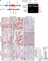
Figure 1.
Figure 1.
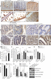
Targeted gene deletion of Fbxw7 in mouse intestine. (A) Schematic shows the floxed Fbxw7 allele (Fbxw7fl/fl) before and after Cre recombination to generate gut-specific conditional Fbxw7 inactivation (Fbxw7ΔG) mice. Arrows indicate location of PCR primers (black, LoxP; and red, Fbxw7) used. (B) PCR analysis of genomic DNA from brain (Br), heart (Hr), and intestine (In) from Fbxw7fl/fl and Fbxw7ΔG mice. OLF-F7/OLR-DF7 PCR shows Fbxw7 gene status. HPRT was used as a loading control. (C–H) IHC for Fbxw7 on representative intestinal tissue sections of 6-wk-old Fbxw7fl/fl (C and D), Fbxw7ΔG/fl (E and F), and Fbxw7ΔG (G and H) mice. Figures are shown at low magnification (C, E, and G) and at high magnification (D, F, and H). Red arrowheads show cells with nuclear Fbxw7, and black arrowheads show cells with cytoplasmic Fbxw7. Experiments were performed for each genotype and for all four different sections (jejunum, ileum, cecum, and colon) and repeated on at least three independent occasions. (I–K) ISH for Fbxw7 on representative intestinal tissue sections of 6-wk-old Fbxw7fl/fl (I), Fbxw7ΔG/fl (J), and Fbxw7ΔG (K) mice. In I and J, yellow arrowheads indicate Fbxw7+ cells in villi, and black arrowheads indicate Fbxw7+ cells in crypts. Boxes indicate magnified intestinal crypts. Experiments were performed in duplicate for each genotype and repeated on at least two independent occasions. (L) qRT-PCR analysis of Fbxw7 mRNA expression in crypts and villi isolated from Fbxw7fl/fl intestine and Fbxw7fl/fl, Fbxw7ΔG/fl, and Fbxw7ΔG whole intestines. Data are mean ± SEM (n = 3; **, P < 0.01; ***, P < 0.001). Experiments were performed in triplicate for each genotype and repeated at least on three independent occasions. Bars: (C, E, G, and I–K) 50 µm; (D, F, and H) 10 µm.
Figure 2.
Figure 2.
In the ApcMin/+ mouse, gut-specific conditional Fbxw7 inactivation (ApcMin/+Fbxw7ΔG mice) induced tumorigenesis and compromised lifespan and survival. (A and B) H&E staining (A) and IHC for β-catenin (B) on duodenum from 3-wk-old ApcMin/+Fbxw7ΔG mice. Experiments were performed in duplicate for all four different sections for each genotype and repeated on at least two independent occasions. Dashed lines indicate intestinal polyps and adenomas (A, green) expressing high amounts of β-catenin (B, black). Bars, 100 µm. (C) Kaplan-Meier survival curves for Fbxw7ΔG, ApcMin/+Fbxw7fl/fl, and ApcMin/+Fbxw7ΔG mice showing the percentage of mice remaining alive at 1-wk intervals. The x axis indicates times (weeks) until mice were euthanized because of severe weight loss. Groups of 14 mice per genotype were used for analysis, and survival curves were statistically different from each other (P < 0.001). (D) β-Catenin polyps (graph shows mean ± SEM) were counted in ApcMin/+Fbxw7fl/fl and ApcMin/+Fbxw7ΔG mice (n = 4 of each genotype according to intestinal locations) on two independent occasions.
Figure 3.
Figure 3.
Protein expression of Fbxw7 substrates and mRNA levels of genes responsible for intestinal differentiation, proliferation, and progenitor markers are altered in Fbxw7ΔG intestinal ECs. (A) The mean percentage of caspase-3+ cells (±SEM) in crypts of Fbxw7fl/fl and Fbxw7ΔG mice (n = 3 for each genotype; 200 crypts each; ***, P < 0.001). (B) Ki67-stained cells in crypts of Fbxw7fl/fl and Fbxw7ΔG mice (n = 3 for each genotype; 100 crypts each) according to intestinal locations. Data present mean per crypt ± SEM (n = 3; ***, P < 0.001). (C) Western blot analysis of Fbxw7fl/fl and Fbxw7ΔG intestinal proteins using antibodies against Fbxw7, Notch 1, Notch 4, p–c-Jun, p–c-Myc, cyclin E, and the loading control β-actin. Experiments were performed on at least two independent occasions. IB, immunoblot. (D and E) qRT-PCR analysis of mRNA encoding the indicated genes responsible for intestinal differentiation, proliferation, and marking progenitors in Fbxw7ΔG and Fbxw7fl/fl intestine. Results were normalized to β-actin in the same sample, and data are presented as fold induction/repression over Fbxw7fl/fl mice. Mean ± SD (n = 3; *, P < 0.05; **, P < 0.01; ***, P < 0.001). Experiments were performed in triplicate for each genotype and repeated on at least three independent occasions.
Figure 4.
Figure 4.
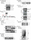
Expression of DEK and TPM is altered in the intestine of Fbxw7ΔG mice. (A–H) Two-dimensional gel/MS-based protein identification using mouse intestinal proteins fractionated into cytosolic (A, Fbxw7fl/fl; C, Fbxw7ΔG) and nuclear extracts (E, Fbxw7fl/fl; G, Fbxw7ΔG). Circled areas in A, C, E, and G are magnified and shown in B, D, F, and H. Blue and yellow circles (B and D) denote isoforms of Tpm; the red circle (H) denotes DEK. (I–L) IHC for TPM and DEK on representative intestinal tissues of 6-wk-old Fbxw7fl/fl (I and K) and Fbxw7ΔG mice (J and L). Dashed lines indicate the boundary of the muscle and epithelia. Arrowheads denote DEK-expressing cells. Bars, 50 µm. (M) Western blot analysis of TPMs in epithelia-enriched and whole intestine protein samples from Fbxw7fl/fl and Fbxw7ΔG mice. Arrows indicate possible transition of TPM isoforms. (N) Western blot analysis of Fbxw7fl/fl and Fbxw7ΔG intestines with DEK, Muc2, and β-actin (loading control) antibodies. (O) qRT-PCR analysis of DEK mRNA in the intestine of Fbxw7ΔG and Fbxw7fl/fl mice. Results were normalized to β-actin expression in the same sample, and data are presented as fold over Fbxw7fl/fl mice (mean ± SD; n = 3; ***, P < 0.001). Experiments were performed in triplicate for each genotype and repeated on at least three independent occasions.
Figure 5.
Figure 5.
TPM abolishment in epithelium and DEK accumulation in crypt of ApcMin/+Fbxw7ΔG mice and human colorectal tumors. (A–D) The expression pattern of DEK and TPM of day 3 postnatal ApcMin/+Fbxw7fl/fl (A and C) and ApcMin/+Fbxw7ΔG (B and D) intestinal tissues. Dashed lines indicate the boundary of the muscle and epithelia. (E–N) IHC for TPM and DEK on representative human colorectal tissues. (E and F) IHC for TPM and DEK in representative sections of normal human colorectal tissues. (G–N) IHC for TPM and DEK in sections of human colorectal tumors with and without FBXW7 mutations. H, J, L, and N show magnified versions of the boxed regions in G, I, K, and M. (A and H) Arrowheads indicate TPM+ cells. SM, smooth muscle cell. (O) HCT116 human CRC cells expressing the indicated FBXW7 genotypes were cultured in vitro. Graph shows mean ± SEM, and numbers in parentheses indicate the number of colonies generated from 50 cells seeded in 24-well dishes in triplicate and on two independent experiments. (P–R) FLAG–TPM1-α or control vector (pcDNA) was expressed in HCT116+/+ and HCT116−/− cells, and status was monitored for cell cycle via flow cytometry and propidium iodide staining (P). Wound healing (Q) and cell migration (R) were also performed. For quantification of colony area and wound healing, images of wounds and colonies were converted into binary images using the ImageJ program. (S) Cell cycle status of HCT116 cells transfected with expression vector pGFP-DEK (HCT116GFP-DEK), DEK-specific duplex siRNA (HCT116DEK−), and sequence scrambled control siRNA (HCT116ssc) was monitored via flow cytometry and propidium iodide staining in triplicates. (O–S) Error bars present mean ± SEM of three independent experiments (*, P < 0.05; **, P < 0.01; ***P < 0.001). (T) DEK, FBXW7, GSK-3β, and β-catenin expression was measured by Western blotting. Bars: (A–G, I, K, and M) 50 µm; (H, J, L, and N) 10 µm.
Figure 6.
Figure 6.
DEK is regulated by the SCF E3 ligase containing the FBXW7-α subunit in a manner dependent on GSK-3β. (A) FLAG-tagged versions of the indicated proteins were expressed in HEK293T cells, and lysates were subjected to immunoblotting with anti-FLAG and anti-GFP. β-Actin was used as a loading control. (B) 35S-labeled DEK was generated by IVT using rabbit RT lysates, with or without λ-phosphatase (λPPase) treatment. Schematic shows potential GSK-3β phosphorylation sites on DEK; red letters document residues mutated in DEK2A. SE, short exposure; LE, long exposure. (C and D) Mutant and wild-type versions of DEK together with other indicated constructs were expressed in HEK293 cells. Cells were treated or not with LiCl and/or proteasome inhibitor I (Prot Inhib) for 8 h (C) or cycloheximide (CHX; D) for the indicated time points. Lysates were examined by Western blotting. (E) HCT116 cells lacking or expressing FBXW7 were transfected with the indicated constructs, labeled with [35S]methionine, and chased with cold methionine for the indicated time points. Lysates were subjected to IP and immunoblotting with anti-GFP. (F) HEK293T cells were transfected with the indicated constructs and subjected to IP and immunoblotting with the indicated antibodies. The asterisk indicates a nonspecific band. (G) FLAG-FBXW7 was generated by IVT, and interaction with DEK-GFP was probed by IP and immunoblotting with the indicated antibodies. (H) HEK293T cells expressing the indicated constructs and HA-tagged ubiquitin (HA-Ub) were subjected to IP and immunoblotting with the indicated antibodies. Experiments in B and E–H and A, C, and D were repeated at least two and three times, respectively.
Figure 7.
Figure 7.
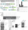
GSK-3β–p-DEK regulates TPM alternative splicing. (A) Schematic of bicistronic reporter construct, TN24tm, used for splicing test. Tpm splicing sites are uppercase and introns are lowercase letters. Splicing and nonsplicing patterns of β-gal and Luc proteins are shown in boxes. Arrows indicate the location of primers for RT-PCR analyses. Red letters indicate stop codons. Ad, adenovirus; pA, poly A signal; SK, skeletal muscle. (B) TN24tm splicing activity assessed by Luc and β-gal expression in HCT116 cells expressing the indicated constructs. Histogram shows the ratio of Luc/β-gal expression (mean ± SEM from three independent experiments). (C) RT-PCR analyses of TN24tm splicing efficiency using RNA from HCT116 cells cotransfected with the indicated constructs. (D) HCT116 cells lacking or expressing FBXW7 were subjected to immunoblotting with the indicated antibodies. The arrowhead marks a higher molecular weight form of TPM. (E) TN24tm splicing assays in HCT116−/− cells transfected with DEK-specific duplex siRNA or scrambled control siRNA. Left panel presents the ratio of Luc/β-gal expression (mean ± SEM from three independent experiments). Middle panel represents the RT-PCR analysis. Right panel shows Western blotting with the indicated antibodies. Arrowheads mark a higher and lower molecular weight form of TPM. The asterisk indicaets a nonspecific band.
Figure 8.
Figure 8.
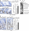
Accumulation of proliferating cells in the crypts of ApcMin/+Fbxw7ΔG mice. (A and B) IHC for Ki67 expression (left) and quantification of Ki67+ cells (right) from intestines of day 3 postpartum mice (A) or from crypts of 3-wk-old mice (B; >200 crypts in each mouse; n = 3 of each genotype). Error bars represent mean ± SEM (***, P < 0.001). Bars, 50 µm. (C) Protein extracts from small intestine of 3-d-postpartum mice of indicated genotypes were analyzed by Western blotting. The asterisk indicates a nonspecific band. The experiment was repeated twice.

V体育官网 - References

    1. Akiyoshi T., Nakamura M., Yanai K., Nagai S., Wada J., Koga K., Nakashima H., Sato N., Tanaka M., Katano M. 2008. Gamma-secretase inhibitors enhance taxane-induced mitotic arrest and apoptosis in colon cancer cells. Gastroenterology. 134:131–144 10.1053/j.gastro.2007.10.008 - DOI - PubMed
    1. Alexiadis V., Waldmann T., Andersen J., Mann M., Knippers R., Gruss C. 2000. The protein encoded by the proto-oncogene DEK changes the topology of chromatin and reduces the efficiency of DNA replication in a chromatin-specific manner. Genes Dev. 14:1308–1312 - "V体育ios版" PMC - PubMed
    1. Allen A., Hutton D.A., Pearson J.P. 1998. The MUC2 gene product: a human intestinal mucin. Int. J. Biochem. Cell Biol. 30:797–801 10.1016/S1357-2725(98)00028-4 - DOI - PubMed
    1. Bharadwaj S., Prasad G.L. 2002. Tropomyosin-1, a novel suppressor of cellular transformation is downregulated by promoter methylation in cancer cells. Cancer Lett. 183:205–213 10.1016/S0304-3835(02)00119-2 - VSports在线直播 - DOI - PubMed
    1. Bonetti P., Davoli T., Sironi C., Amati B., Pelicci P.G., Colombo E. 2008. Nucleophosmin and its AML-associated mutant regulate c-Myc turnover through Fbw7 γ. J. Cell Biol. 182:19–26 10.1083/jcb.200711040 - VSports - DOI - PMC - PubMed

Publication types

"VSports app下载" MeSH terms