V体育2025版 - Skip to main page content
U.S. flag

An official website of the United States government

Dot gov

The . gov means it’s official. Federal government websites often end in VSports app下载. gov or . mil. Before sharing sensitive information, make sure you’re on a federal government site. .

Https

The site is secure V体育官网. The https:// ensures that you are connecting to the official website and that any information you provide is encrypted and transmitted securely. .

. 2011 Feb;121(2):800-8.
doi: 10.1172/JCI43114. Epub 2011 Jan 25.

Notch1 loss of heterozygosity causes vascular tumors and lethal hemorrhage in mice

Affiliations

Notch1 loss of heterozygosity causes vascular tumors and lethal hemorrhage in mice

Zhenyi Liu et al. J Clin Invest. 2011 Feb.

Abstract

The role of the Notch signaling pathway in tumor development is complex, with Notch1 functioning either as an oncogene or as a tumor suppressor in a context-dependent manner. To further define the role of Notch1 in tumor development, we systematically surveyed for tumor suppressor activity of Notch1 in vivo. We combined the previously described Notch1 intramembrane proteolysis-Cre (Nip1::Cre) allele with a floxed Notch1 allele to create a mouse model for sporadic, low-frequency loss of Notch1 heterozygosity. Through this approach, we determined the cell types most affected by Notch1 loss. We report that the loss of Notch1 caused widespread vascular tumors and organism lethality secondary to massive hemorrhage VSports手机版. These findings reflected a cell-autonomous role for Notch1 in suppressing neoplasia in the vascular system and provide a model by which to explore the mechanism of neoplastic transformation of endothelial cells. Importantly, these results raise concerns regarding the safety of chronic application of drugs targeting the Notch pathway, specifically those targeting Notch1, because of mechanism-based toxicity in the endothelium. Our strategy also can be broadly applied to induce sporadic in vivo loss of heterozygosity of any conditional alleles in progenitors that experience Notch1 activation. .


"VSports" Figures

Figure 1
Figure 1. N1::Cre excises floxed Notch1 after activation.
(A and B) Loss of functional Notch1 is a low-frequency event, since only a small number of Cre6MT molecules reach the nucleus. C-terminal 6MT is omitted from the diagram for clarity. CoA, coactivator; CoR, corepressor; MAM, Mastermind. (C) All N1::Crelo/fl mice developed corneal plaques after 2–3 months, and this phenotype became more severe as animals aged. LacZ staining revealed that the transparent corneal was transformed into a skin-like structure with neoangiogenesis. Not all ECs and keratinocytes were labeled with lacZ. (D) N1::Crelo/fl mice showed increased angiogenesis in the ear. Scale bars: 150 μm (C); 10 mm (D).
Figure 2
Figure 2. N1::Crelo/fl mice developed vascular tumors.
(A) Kaplan-Meier survival analysis of 41 mutant and 45 control animals. Animals euthanized or alive at the time of analysis were censored (44 control and 26 mutant). (B) Summary of the major pathological findings from a comprehensive analysis of 13 pairs of control and mutant animals. Some animals developed hemangiomas in multiple tissues, making the tumor count greater than 13. The distribution of hemangiomas in different organs is further dissected (see Supplemental Table 1). (CF) Representative images of spleen (C) and liver from N1::Crelo/+ (D) and N1::Crelo/fl animals (E and F). (E) Liver from a naturally deceased animal. (F) Liver from a euthanized N1::Crelo/fl animal, in which the tumors were still intact and filled with blood. (GI) H&E liver sections from N1::Crelo/+ (G) and N1::Crelo/fl animals (H and I). See Results for details. (JL) Immunostaining of liver tumor for PECAM (J), vWF (K), and PECAM and Ki67 (L). Inset in L shows the enlarged view of boxed area. Representative vascular tumors in other organs are shown in M (skin; H&E) and N (ovary; vWF). Scale bars: 50 μm; 10 μm (L, inset).
Figure 3
Figure 3. Imaging tumor progression.
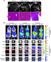
(A) MRI was used to assess tumor progression in the liver. H&E images of the liver from respective animals are also shown. Scale bar: 100 μm. (B) Bioluminescence imaging of live animals and freshly dissected organs at the specified ages. Shown are mean and SD of organ luciferase signal (105 photons/s) from at least 4 different individuals of the same genotype. Organs showing statistically significant differences in signal intensity are boxed in red. See Supplemental Figure 6 for images of more organs.
Figure 4
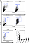
Figure 4. Comparison of the RosaEYFP reporter labeling efficiency in ECs of different organs in N1::Crelo/+RosaEYFP/LacZ mice.
(A) ECs from liver, kidney, spleen, heart, and lung were enriched, stained with CD45 and CD31, and subjected to flow cytometry analysis. Percent CD31+EYFP and CD31+EYFP+ cells among CD45 cells is indicated. (B) Statistical analysis on 4 N1::Crelo/+RosaEYFP/LacZ mice showed that the liver had the highest percentage of EYFP+ ECs (about 20%, compared with less than 5% in other organs). *P < 0.05.
Figure 5
Figure 5. Characterization of vascular tumor cells.
(A) LacZ-, PECAM/EYFP-, and BrdU/EYFP-stained liver sections. A single, 2-hour-long pulse of BrdU labeled 5%–10% of ECs. Inset in right panel shows the enlarged view of boxed area. Scale bars: 50 μm; 10 μm (inset). (BE) Immunostaining of phospho-Rb. Phospho-Rb signal was absent in areas without tumor (B and C); the presence of the signal (brown) is shown in D and E. Sections were counterstained with hematoxylin. The tumor identity in D and E was confirmed with PECAM and Ki67 costaining in Figure 2L (adjacent sections). Boxed regions in B and D are shown at higher magnification in C and E, respectively. Scale bars: 100 μm.
Figure 6
Figure 6. Loss of Notch1 in ECs is associated with their rapid expansion.
(A) Liver ECs were sorted to remove CD45+ cells and then separated based on the expression of EYFP and either CD105 (proliferating EC marker) or CD31 (pan-EC marker). (B) PCR analysis confirmed the deletion of floxed Notch1 allele in the majority of CD45CD105+EYFP+ and CD45CD105+EYFP cells; in contrast, a substantial amount of floxed alleles was still detected in CD45CD31+EYFP+ and CD45CD31+EYFP cells. The primers bind at positions illustrated in the diagram. Lanes a–f correspond with gates in A. M, molecular marker.

Comment in

References

    1. Kopan R, Ilagan MX. The canonical Notch signaling pathway: unfolding the activation mechanism. Cell. 2009;137(2):216–233. doi: 10.1016/j.cell.2009.03.045. - DOI - PMC - PubMed
    1. Koch U, Radtke F. Notch and cancer: a double-edged sword. Cell Mol Life Sci. 2007;64(21):2746–2762. doi: 10.1007/s00018-007-7164-1. - DOI - PMC - PubMed
    1. Weng AP, et al. Activating mutations of NOTCH1 in human T cell acute lymphoblastic leukemia. Science. 2004;306(5694):269–271. doi: 10.1126/science.1102160. - DOI - PubMed
    1. Jeannet R, et al. Oncogenic activation of the Notch1 gene by deletion of its promoter in Ikaros-deficient T-ALL [published online ahead of print September 9, 2010].Blood . doi: 10.1182/blood-2010-05-286658. - PMC - PubMed
    1. Demehri S, Turkoz A, Kopan R. Epidermal Notch1 loss promotes skin tumorigenesis by impacting the stromal microenvironment. Cancer Cell. 2009;16(1):55–66. doi: 10.1016/j.ccr.2009.05.016. - DOI (V体育ios版) - PMC - PubMed

VSports app下载 - Publication types

V体育ios版 - MeSH terms

Substances