<u dropzone="fmYhjS"></u>
VSports - Skip to main page content
U.S. flag

An official website of the United States government

Dot gov

The . gov means it’s official. Federal government websites often end in VSports app下载. gov or . mil. Before sharing sensitive information, make sure you’re on a federal government site. .

Https

The site is secure V体育官网. The https:// ensures that you are connecting to the official website and that any information you provide is encrypted and transmitted securely. .

. 2011 Mar 1;71(5):1730-41.
doi: 10.1158/0008-5472.CAN-10-1432. Epub 2011 Jan 21.

"V体育2025版" An EGFR-Src-Arg-cortactin pathway mediates functional maturation of invadopodia and breast cancer cell invasion

Affiliations

An EGFR-Src-Arg-cortactin pathway mediates functional maturation of invadopodia and breast cancer cell invasion

Christopher C Mader et al. Cancer Res. .

"VSports" Abstract

Invasive carcinoma cells use specialized actin polymerization-driven protrusions called invadopodia to degrade and possibly invade through the extracellular matrix (ECM) during metastasis VSports手机版. Phosphorylation of the invadopodium protein cortactin is a master switch that activates invadopodium maturation and function. Cortactin was originally identified as a hyperphosphorylated protein in v-Src-transformed cells, but the kinase or kinases that are directly responsible for cortactin phosphorylation in invadopodia remain unknown. In this study, we provide evidence that the Abl-related nonreceptor tyrosine kinase Arg mediates epidermal growth factor (EGF)-induced cortactin phosphorylation, triggering actin polymerization in invadopodia, ECM degradation, and matrix proteolysis-dependent tumor cell invasion. Both Src and Arg localize to invadopodia and are required for EGF-induced actin polymerization. Notably, Arg overexpression in Src knockdown cells can partially rescue actin polymerization in invadopodia while Src overexpression cannot compensate for loss of Arg, arguing that Src indirectly regulates invadopodium maturation through Arg activation. Our findings suggest a novel mechanism by which an EGFR-Src-Arg-cortactin pathway mediates functional maturation of invadopodia and breast cancer cell invasion. Furthermore, they identify Arg as a novel mediator of invadopodia function and a candidate therapeutic target to inhibit tumor invasion in vivo. .


Figures

Figure 1
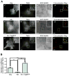
Figure 1. Arg and Src, but not Abl, localize to invadopodia
(A) MDA-MB-231 cells were plated on Alexa-405 labeled gelatin matrix, fixed, and labeled for Arg (a1) or Abl (a2), and the invadopodia marker, Tks5 (a2 and b2). Cells expressing Src-TagRFP and knocked down for endogenous Src (c1) were plated on Alexa-405 gelatin, fixed, and labeled for Tks5 as above (c2). Co-localization masks are shown for the kinases with Tks5 at invadopodia actively degrading Alexa-405 gelatin matrix (a3,4, b3,4, c3,4). (B) Quantification of the co-localization (Manders coefficient) between Arg, Abl, Src, and Tks5 at matrix-degrading invadopodia. n=24 (Arg), n=17 (Abl), n=13 (Src). Scale bars=10 μm.
Figure 2
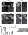
Figure 2. Arg is required for cortactin phosphorylation at invadopodium precursors
(A) MDA-MB-231 cells stably expressing cortactin-TagRFP were treated with siRNA specific for control, Arg, Abl, or Src, plated on fibronectin/gelatin matrix and starved before EGF stimulation. Cells were fixed and labeled for phosphorylated cortactin using a phosphospecific cortactin antibody (anti-pY421) before (0min.) or after (3min.) EGF stimulation. (B) Cortactin phosphorylation was measured as the ratio of pY421 signal/cortactin-TagRFP signal at invadopodium precursors. Control siRNA: n=105(0min.)/n=127(3min.), Abl siRNA: n=110(0min.)/n=97(3min.), Arg siRNA: n=138(0min.)/n=136(3min.), Src siRNA: n=122(0min.)/n=160(3min.). Scale bars=10 μm. (C) MDA-MB-231 cells were treated with increasing concentrations of EGF for 3 minutes, lysed, immunoprecipitated with anti-cortactin antibodies followed by immunoblotting with anti-Arg antibodies. The samples were blotted with anti-cortactin as control.
Figure 3
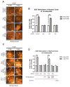
Figure 3. Arg kinase activity is required for actin barbed end generation at invadopodia
(A) MDA-MB-231 cells were knocked down using control, Arg, Abl, or Src siRNA and either left untreated (a,c,e,g) or stimulated with EGF for 3 minutes (b,d,f,h). Cells were fixed and labeled for biotin-actin and Arp2 as an invadopodium marker. (B) Quantification of free actin barbed ends as measured by average biotin-actin intensity in stimulated invadopodia containing Arp2. Control siRNA: n=404(0min.)/n=365(3min.), Abl siRNA: n=312(0min.)/n=271(3min.), Arg siRNA: n=283(0min.)/n=252(3min.), Src siRNA: n=296(0min.)/n=256(3min.). (C) Cells stably expressing RNAi-resistant rescue mutants of Arg (Arg-YFP or Arg KI-YFP=kinase inactive) or YFP alone were knocked down using Arg siRNA, stimulated and labeled as in (A) (i-n). Alternatively, MDA-MB-231 cells were treated with 3.3 μM STI-571 prior to EGF stimulation (o, p). (D) Quantification of free actin barbed ends from experiments in (C). Arg-YFP: n=308(0min.)/n=209(3min.), YFP: n=127(0min.)/n=275(3min.), ArgKI-YFP: n=196(0min.)/n=181(3min.), STI-571: n=289(0min.)/n=415(3min.). Scale bars=5μm.
Figure 4
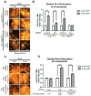
Figure 4. Arg is required for Src-mediated stimulation of actin barbed end formation in invadopodia
(A) Representative images of MDA-MB-231 cells stably expressing Src-TagRFP (a-d) or cells stably expressing Arg-YFP (e-h), treated with control siRNA (a,b,e,f), Arg siRNA (c,d), or Src siRNA (g,h). Cells were either left untreated (a,c,e,g), or stimulated with EGF for 3 min. (b,d,f,h), fixed and stained for biotin-actin and Arp2 as an invadopodium marker. (B) Quantification of barbed ends as measured by the average actin intensity at stimulated invadopodia in response to EGF (all conditions n=200). The difference between all 3 minute stimulation values are statistically significant as measured by individual Post-Hoc student’s t-test (p<0.01). Scale bars=5μm. (C) Representative images of cells knocked down for endogenous Arg and re-expressing YFP, Arg-YFP, or mutant Arg Y439F-YFP. Scale bars=5μm. (D) Cells were stimulated and treated as above, and actin barbed ends were quantified as in B. YFP: n=127(0min.)/n=275(3min.), Arg-YFP: n=308(0min.)/n=209(3min.), ArgKI-YFP: n=196(0min.)/n=181(3min.), STI-571: n=289(0min.)/n=415(3min.).
Figure 5
Figure 5. Arg is required for Src-mediated cortactin phosphorylation in invadopodia
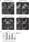
(A) MDA-MB-231 cells stably expressing cortactin-TagRFP were treated with control siRNA, Arg siRNA, or Src siRNA and transfected with either Arg-YFP (two upper panels) or Src-YFP (two lower panels), plated on fibronectin/gelatin matrix, and starved before EGF stimulation. The cells were fixed and labeled for phosphorylated cortactin using a phosphospecific cortactin antibody (anti-pY421) before (0min.) or after (3min.) EGF stimulation. Scale bars=20μm. (B) Cortactin phosphorylation was measured as the ratio of pY421 signal/cortactin-TagRFP signal at invadopodium precursors. Control siRNA+ArgYFP: n=50(0min.)/n=49(3min.), Src siRNA+ArgYFP: n=48(0min.)/n=53(3min.), Control siRNA+SrcYFP: n=55(0min.)/n=48(3min.), Arg siRNA+SrcYFP: n=48(0min.)/n=48(3min.).
Figure 6
Figure 6. Arg kinase activity is required for extracellular matrix degradation and invasion
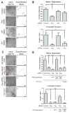
(A) MDA-MB-231 cells treated with control, Arg, Abl, or Src siRNA were plated on Alexa-568 fibronectin/gelatin matrix and allowed to degrade for 22 hours. Shown are representative images (left panels) and quantification masks (right panels) of degradation areas. (B) Quantification of matrix degradation from conditions as in A; n=60 fields (upper graph) and MDA-MB-231 cells knocked down as in A and B were plated on Matrigel-coated membranes and allowed to invade for 20 hours; n=12 (lower graph) (C) MDA-MB-231 cells stably expressing Arg-YFP, Arg KI-YFP, or YFP control were treated with Arg siRNA, control siRNA, or with STI-571, and plated on fibronectin/gelatin matrix and allowed to degrade as in A. Shown are representative images (left panels) and quantification masks (right panels). (D) Quantification of matrix degradation area from conditions in C. YFP n=31, Arg-YFP n=30, ArgKI-YFP n=30, STI-571 n=10. MDA-MB-231 cells were plated on Matrigel-coated membranes and allowed to invade, as described in C. YFP n=8, Arg-YFP n=6, ArgKI-YFP n=8. Invasion was normalized to migration rate, proteolysis-independent invasion, and protein re-expression levels where appropriate. Scale bars=10μm.
Figure 7
Figure 7. Model for EGF-stimulated activation of actin polymerization at invadopodia
(A) Cortactin and actin form invadopodium precursors, to which Arg is recruited via a phosphorylation-independent binding interaction. (B) EGF stimulation promotes dimerization and tyrosine phosphorylation of the receptor. The phosphorylated receptor binds to the Arg SH2 domain, allowing for autophosphorylation of its linker tyrosine (Y272) and weak activation. The phosphorylated receptor also binds and activates Src kinase, which can phosphorylate Arg on its activation loop tyrosine (Y439), resulting in full Arg kinase activation. (C) Fully activated Arg phosphorylates cortactin at invadopodium precursors. (D) Phosphorylated cortactin induces Arp2/3-dependent actin polymerization. NTA, N-terminal acidic region; VCA, verprolin-cofilin-acidic domain; PolyP, poly-proline domain.

References (VSports最新版本)

    1. Barsky SH, Siegal GP, Jannotta F, Liotta LA. Loss of basement membrane components by invasive tumors but not by their benign counterparts. Lab Invest. 1983;49:140–7. - PubMed
    1. Yamaguchi H, Wyckoff J, Condeelis J. Cell migration in tumors. Curr Opin Cell Biol. 2005;17:559–64. - PubMed
    1. Wyckoff J, Wang W, Lin EY, et al. A paracrine loop between tumor cells and macrophages is required for tumor cell migration in mammary tumors. Cancer Res. 2004;64:7022–9. - PubMed
    1. Gimona M, Buccione R, Courtneidge SA, Linder S. Assembly and biological role of podosomes and invadopodia. Curr Opin Cell Biol. 2008;20:235–41. - PubMed
    1. Linder S. Invadosomes at a glance. J Cell Sci. 2009;122:3009–13. - PubMed

Publication types (V体育平台登录)

MeSH terms (V体育官网)