Skip to main page content
U.S. flag

An official website of the United States government

Dot gov

The . gov means it’s official VSports app下载. Federal government websites often end in . gov or . mil. Before sharing sensitive information, make sure you’re on a federal government site. .

Https

The site is secure V体育官网. The https:// ensures that you are connecting to the official website and that any information you provide is encrypted and transmitted securely. .

. 2011 Mar 18;286(11):9587-97.
doi: 10.1074/jbc.M110.202911. Epub 2011 Jan 12.

Autophagy controls IL-1beta secretion by targeting pro-IL-1beta for degradation

Affiliations

"V体育官网入口" Autophagy controls IL-1beta secretion by targeting pro-IL-1beta for degradation

James Harris et al. J Biol Chem. .

Abstract

Autophagy is a key regulator of cellular homeostasis that can be activated by pathogen-associated molecules and recently has been shown to influence IL-1β secretion by macrophages. However, the mechanisms behind this are unclear. Here, we describe a novel role for autophagy in regulating the production of IL-1β in antigen-presenting cells. After treatment of macrophages with Toll-like receptor ligands, pro-IL-1β was specifically sequestered into autophagosomes, whereas further activation of autophagy with rapamycin induced the degradation of pro-IL-1β and blocked secretion of the mature cytokine. Inhibition of autophagy promoted the processing and secretion of IL-1β by antigen-presenting cells in an NLRP3- and TRIF-dependent manner. This effect was reduced by inhibition of reactive oxygen species but was independent of NOX2. Induction of autophagy in mice in vivo with rapamycin reduced serum levels of IL-1β in response to challenge with LPS. These data demonstrate that autophagy controls the production of IL-1β through at least two separate mechanisms: by targeting pro-IL-1β for lysosomal degradation and by regulating activation of the NLRP3 inflammasome. VSports手机版.


Figures
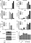
FIGURE 1.
FIGURE 1.
The autophagy inhibitors 3-MA and wortmannin induce endotoxin-dependent inflammasome activation. A, iBMM were treated with LPS (100 ng/ml) and 3-MA at the concentrations indicated for 24 h, and supernatants were analyzed for IL-1β, IL-6, and TNF-α by ELISA. B, BMDC were treated with LPS (10 ng/ml) and wortmannin or 3-MA at the concentrations indicated for 24 h, and supernatants were analyzed for IL-1β by ELISA. C, BMDC were treated with LPS (10 ng/ml) and 3-MA at the concentrations indicated for 24 h, and supernatants were analyzed for IL-1α by ELISA. D, Western blot analysis of IL-1β in cell lysates and supernatants from BMDC stimulated with LPS (10 ng/ml) and 3-MA (10 mm) for 24 h (representative of three separate experiments). E, ELISA analysis of IL-18 from supernatants of iBMM stimulated with LPS (100 ng/ml) and the indicated concentrations of 3MA for 24 h. Data are means ± S.D. from one experiment representative of 2 (A, C, and E) or 3 (B). *, p < 0.05, significant difference from cells treated with LPS alone (one-way ANOVA, followed by Tukey's HSD test).
FIGURE 2.
FIGURE 2.
3-MA activates the inflammasome through inhibition of type III PI3K and autophagy. A, Western blot analysis of LC3 in cell lysates from iBMM treated with LPS (100 ng/ml) or poly(I:C) (p(I:C), 10 μg/ml) and 3-MA (10 nm) for 6 h (representative blot from three separate experiments). Cells were treated with or without bafilomycin A (baf; 100 nm). B, densitometric measurement of the ratio of LC3 II:β-actin staining in A. C, confocal analysis of GFP-LC3-expressing iBMM treated with LPS (100 ng/ml) and 3-MA (10 mm) for 6 h. D, ELISA analysis of IL-1β in the supernatants of iBMM transfected with siRNA against beclin 1 (Bec1) or control (scr) siRNA and treated with different concentrations of LPS. Bec1 protein knockdown was confirmed by Western blot (inset). E, BMDC were treated with LPS (10 ng/ml) and either wortmannin (wort), the PI3KIα inhibitor 2,3-[4-(4-morpholinyl)thieno[3,2-d]pyrimidin-2-yl]-phenol or the PI3kIδ inhibitor IC87114 for 24 h, and supernatants were analyzed for IL-1β by ELISA. F, BMDC were treated with LPS (10 ng/ml) and Akt inhibitor (10 μm) for 24 h, and IL-1β was measured in the supernatants by ELISA. G, BMDC were treated with LPS (10 ng/ml) with ATP (5 mm), alum (5 mm), or 3-MA for 24 h. Cells were stained with PI and analyzed by flow cytometry. Data are means ± S.D. from one experiment representative of two (D, E, and F) or mean ± S.E. from three (G) experiments. *, p < 0.05, significant difference from cells treated with TLR agonist alone (one-way ANOVA, followed by Tukey's HSD test).
FIGURE 3.
FIGURE 3.
3-MA-induced inflammasome activation is dependent on NLRP3. A, BMDC from WT C57Bl/6 and NLRP3−/− mice were treated with PAM3CysK4 (PAM; 1 μg/ml), poly(I:C) (p(I:C); 10 μg/ml), LPS (10 ng/ml), or CpG (10 μg/ml) with 3-MA (10 mm) for 24 h, and supernatants were analyzed for IL-1β by ELISA. WT and caspase 1−/− (B) or ASC/ (C) iBMM were treated with LPS (100 ng/ml) and indicated concentrations of 3MA for 24 h, and supernatants were analyzed by ELISA for IL-1β secretion. D, BMDC from WT C57Bl/6, MyD88/, and TRIF/ mice were treated with LPS (10 ng/ml) plus ATP (5 mm), alum (5 mm), or indicated concentrations of 3-MA for 24 h. Supernatants were analyzed by ELISA for IL-1β. Data are means ± S.D. from one experiment representative of two (B, C, and D) or three (A). *, p < 0.05; significant difference from cells treated with TLR agonist alone (one-way ANOVA, followed by Tukey's HSD test).
FIGURE 4.
FIGURE 4.
3-MA-induced inflammasome activation is dependent on ROS but not NOX2. A, BMDC were treated with LPS (10 ng/ml) and 3-MA (10 mm) with different concentrations of the reactive oxygen species scavenger NAc for 24 h, and supernatants were analyzed by ELISA for IL-1β. B, Western blot analysis of caspase 1 in cell lysates from iBMM treated with LPS (100 ng/ml) and 3-MA (at concentrations indicated) ± NAc (25 mm) for 24 h (representative of two separate experiments). ELISA analysis of IL-1β in supernatants of WT and NOX2/ iBMM treated with LPS (100 ng/ml) plus 3MA (C) or with LPS plus ATP (5 mm) or alum (5 mm) (D) for 24 h. Data are means ± S.D. from one experiment representative of two (C and D) or three (A). *, p < 0.05, significant difference from cells treated with LPS alone (one-way ANOVA, followed by Tukey's HSD test).
FIGURE 5.
FIGURE 5.
Induction of autophagy inhibits IL-1β secretion. A, Western blot analysis of IL-1β in cell lysates from iBMM treated with LPS (100 ng/ml) or PAM3CysK4 (PAM; 1 μg/ml) for 20 h then with rapamycin (rapa; 50 μg/ml) for 4 h (representative of three separate experiments). B, densitometric analysis of the ratio of pro-IL-1β:β-actin staining in A. BMDC were treated with LPS (10 ng/ml) and alum (5 μm) (C) or chitosan (2 μg/ml) (D) with the indicated concentrations of rapamycin and supernatants analyzed for IL-1β by ELISA. E, BMDC were treated with LPS for 20 h, followed by rapamycin (50 μg/ml) for 4 h and ATP (5 mm) for 1 h, and supernatants were analyzed by ELISA for secreted IL-1β and IL-6. Data are means ± S.D. *, p < 0.05, significant difference from cells treated with LPS alone (one-way ANOVA, followed by Tukey's HSD test).
FIGURE 6.
FIGURE 6.
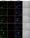
Intracellular IL-1β is targeted by autophagosomes. iBMM stably transfected with GFP-LC3 (green) were treated with LPS (100 ng/ml) for the indicated times, fixed, and stained with antibody against IL-1β (red). Nuclei were stained with bisbenzimide H 33258 (blue). Cells were analyzed by confocal microscopy. Scale bar, 10 μm.
FIGURE 7.
FIGURE 7.
TLR agonists induce targeting of IL-1β by autophagosomes. iBMM stably transfected with GFP-LC3 (green) were treated for 6 h with PAM3CysK4 (PAM; 1 μg/ml), poly(I:C) (p(I:C); 10 μg/ml), R837 (10 μg/ml), or CpG (10 μg/ml). Cells were fixed and stained with antibody against IL-1β (red). Nuclei are stained with bisbenzimide H 33258 (blue). Cells were analyzed by confocal microscopy. Scale bar, 10 μm.
FIGURE 8.
FIGURE 8.
Induction of autophagy reduces IL-1β secretion in vivo. BALB/c mice were injected intraperitoneally with PBS, LPS (10 μg/mouse), or LPS with rapamycin (1.5 mg/kg) and blood taken after 4 h for analysis of IL-1β (A) or IL-12p40 (B) by ELISA. Data are means ± S.E. from a single experiment and analyzed by one-way ANOVA, followed by Tukey's HSD test (n = 5 (PBS and LPS/rapamycin) and n = 4 (LPS alone)).

References

    1. Dinarello C. A. (2010) Eur. J. Immunol. 40, 599–606 - V体育官网 - PubMed
    1. Schroder K., Tschopp J. (2010) Cell 140, 821–832 - PubMed
    1. Eisenbarth S. C., Colegio O. R., O'Connor W., Sutterwala F. S., Flavell R. A. (2008) Nature 453, 1122–1126 - "VSports" PMC - PubMed
    1. Hornung V., Bauernfeind F., Halle A., Samstad E. O., Kono H., Rock K. L., Fitzgerald K. A., Latz E. (2008) Nat. Immunol. 9, 847–856 - PMC - PubMed
    1. Li H., Nookala S., Re F. (2007) J. Immunol. 178, 5271–5276 - PubMed

Publication types

MeSH terms

Substances

LinkOut - more resources