Skip to main page content
U.S. flag

An official website of the United States government

Dot gov

The . gov means it’s official. Federal government websites often end in VSports app下载. gov or . mil. Before sharing sensitive information, make sure you’re on a federal government site. .

Https

The site is secure. The https:// ensures that you are connecting to the official website and that any information you provide is encrypted and transmitted securely V体育官网. .

. 2011 Mar 11;286(10):8308-8324.
doi: 10.1074/jbc.M110.197301. Epub 2010 Dec 20.

Zymophagy, a novel selective autophagy pathway mediated by VMP1-USP9x-p62, prevents pancreatic cell death (V体育ios版)

Affiliations

Zymophagy, a novel selective autophagy pathway mediated by VMP1-USP9x-p62, prevents pancreatic cell death

Daniel Grasso et al. J Biol Chem. .

Abstract

Autophagy has recently elicited significant attention as a mechanism that either protects or promotes cell death, although different autophagy pathways, and the cellular context in which they occur, remain to be elucidated. We report a thorough cellular and biochemical characterization of a novel selective autophagy that works as a protective cell response. This new selective autophagy is activated in pancreatic acinar cells during pancreatitis-induced vesicular transport alteration to sequester and degrade potentially deleterious activated zymogen granules. We have coined the term "zymophagy" to refer to this process VSports手机版. The autophagy-related protein VMP1, the ubiquitin-protease USP9x, and the ubiquitin-binding protein p62 mediate zymophagy. Moreover, VMP1 interacts with USP9x, indicating that there is a close cooperation between the autophagy pathway and the ubiquitin recognition machinery required for selective autophagosome formation. Zymophagy is activated by experimental pancreatitis in genetically engineered mice and cultured pancreatic acinar cells and by acute pancreatitis in humans. Furthermore, zymophagy has pathophysiological relevance by controlling pancreatitis-induced intracellular zymogen activation and helping to prevent cell death. Together, these data reveal a novel selective form of autophagy mediated by the VMP1-USP9x-p62 pathway, as a cellular protective response. .


V体育官网入口 - Figures
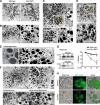
FIGURE 1.
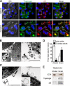
FIGURE 1.
Hyperstimulation of CCK-R in acinar cells induces the redistribution of zymogen granules and VMP1-containing autophagosomes. A–F, transmission electron microscopy of pancreatic tissue. A, wild type and ElaI-VMP1 mouse pancreata. No ultrastructural alteration is evident in zymogen granules of acinar cells from ElaI-VMP1 compared with wild type mouse pancreata. B, wild type mouse pancreas after CCK-R hyperstimulation (detailed on the right). Ultrastructural alterations of zymogen granules (arrowheads), fusion between zymogen granules and condensing vacuoles (arrow), and zymogen granules appearing near basolateral dilated space are shown (*). C–F, ultrastructure of pancreatic tissue from ElaI-VMP1 mice after CCK-R hyperstimulation with cerulein. C and D, autophagosomes containing zymogen granules are observed in the apical area of the acinar cell (dashed line). Zymogen granules inside autophagosomes are detailed in C (bottom panel). Autophagosome-double membrane is detailed in D, bottom panel (arrowhead). E, left panel, example of zymogen granules sequestered by autophagosome (arrow). Right panel, examples of autophagosomes containing one single zymogen granule (arrowheads). F, examples of large autophagosomes and autolysosomes containing zymogen granules. Higher magnification is shown on the right upper panel. At the bottom panel, the arrowheads show the limits of the single membrane autolysosomes (panels a and b) and large double membrane-limited autophagosomes (panels c and d). G, Western blot analysis and densitometry quantification of p62 in ElaI-VMP1 mouse pancreas homogenates shows the progressive reduction of the p62 signal evidencing autophagic flow in pancreas from transgenic mice under CCK-R hyperstimulation with cerulein (**, p < 0.001 versus wild type). H, bright field, VMP1-EGFP fluorescence and merge images of isolated ElaI-VMP1 pancreatic acini. VMP1-EGFP fluorescence is located in the basal region of untreated acini (upper panel). After 1 h of CCK-R hyperstimulation with cerulein, VMP1-EGFP relocated to the granular area of acinar cells (bottom panel). Results are representative of at least three independent experiments. Scale bars, 4 μm (A–F), and 10 μm (H). BL, basolateral, AP, apical, ZG, zymogen granules. N, nucleus.
FIGURE 2.
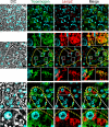
FIGURE 2.
Inducible and selective sequestration of zymogen granules by VMP1-mediated autophagic pathway. A, immunofluorescence assay using anti-LC3 and anti-trypsinogen antibodies on pancreatic acini isolated from ElaI-VMP1 mice. Top row, untreated isolated acini. No colocalization between LC3 and trypsinogen is observed in acinar cells. A detail of granular areas is shown on the upper left side. Bottom row, cerulein-mediated CCK-R hyperstimulated isolated acini. Remarkable colocalization between LC3 and zymogen granules is found in acinar cells. Colocalization examples are detailed. DIC, differential interference contrast. B–D, isolation of autophagosomes using anti-GFP antibody bound magnetic beads from postnuclear supernatant of ElaI-VMP1 mouse pancreas homogenates. B, we show two examples of VMP1-EGFP autophagosomes containing zymogen granules isolated from CCK-R hyperstimulated mouse pancreas homogenates. These double membrane structures containing zymogen granules are detailed in left panel. C, two examples of VMP1-EGFP-coated structures from untreated ElaI-VMP1 mouse pancreas homogenates. Empty or cytoplasm-containing simple or double membrane structures are observed and detailed (right panel). D, quantitative analysis indicates that less than 20% of autophagosomes containing zymogen granules are isolated from untreated animal pancreata, and more than 70% of autophagosomes containing zymogen granules are isolated from CCK-R hyperstimulated mouse pancreata (**, p < 0.001 versus untreated). E, Western blot analysis using anti-LC3, anti-trypsinogen, and anti-p62 antibodies of magnetically immunopurified autophagosomes from CCK-R-hyperstimulated ElaI-VMP1 mouse pancreas homogenates (cerulein lane) compared with autophagosomal fraction obtained from untreated mouse pancreas (untreated lane). The presence of LC3-II (16 kDa) confirms that all the isolated structures are autophagosomes. Zymogen granules as cargo of such autophagosomes are evidenced by trypsinogen-positive blot in autophagosomal fraction isolated from CCK-R-hyperstimulated mouse pancreas. The presence of p62 suggests selective autophagosomes and p62-mediated cargo recognition. Results are representative of at least three independent experiments. Scale bars, 10 μm (A), and 0.5 μm (B and C).
FIGURE 3.
FIGURE 3.
Zymophagy is induced by CCK-R hyperstimulation to selectively sequester and degrade activated zymogen granules. Intracellular activation of trypsinogen in isolated pancreatic acini was detected by BZiPAR reagent (see “Experimental Procedures”). A and B, trypsin activity in isolated pancreatic acini before (top row), 30 min after (middle row), and 1 h after (bottom row) cerulein CCK-R hyperstimulation. Activated trypsin is evidenced by red spots of hydrolyzed substrate. A, acini from wild type mice; early trypsinogen activation is evident within acinar cells during the course of CCK-R hyperstimulation. B, acini from ElaI-VMP1 mice; no activity of trypsin is found after 30 min of CCK-R hyperstimulation, and only few spots of hydrolyzed substrate are detected within acinar cells after 1 h of treatment. EGFP-VMP1 fluorescence shows relocation of VMP1 to the granular area of acinar cells in ElaI-VMP1 pancreatic acini after CCK-R hyperstimulation. C, quantification of trypsin activity in homogenates from isolated pancreatic acini during CCK-R hyperstimulation. Acini from ElaI-VMP1 mice show significantly less trypsinogen activation compared with wild type mice (**, p < 0.001 versus wild type). D, remarkable colocalization between hydrolyzed trypsin substrate (red spots) and VMP1-EGFP fluorescent signals is seen in acinar cells from ElaI-VMP1 mice after 1 h of CCK-R hyperstimulation. Merge image in look-up table (LUT) pseudocolor and two hot fluorescence spots profile are shown to prove specific colocalization. E, trypsinogen activation in ElaI-VMP1 pancreatic acini treated with chloroquine or vinblastine after 1 h of CCK-R hyperstimulation. Red fluorescence spots show increased trypsin activity within acinar cells submitted to both autophagy inhibitory treatments (chloroquine and vinblastine). F, trypsinogen activation was quantified as percentage of maximal trypsin activity in wild type mice pancreatic acini under cerulein CCK-R hyperstimulation. The protective effect of VMP1-autophagy pathway on trypsinogen activation in ElaI-VMP1 acini upon cerulein CCK-R hyperstimulation (ElaI-VMP1 vehicle bar) is significantly reversed by autophagy inhibitors (chloroquine and vinblastine treatment bars); (**, p < 0.001 versus ElaI-VMP1 vehicle bar). No significant differences are observed between vehicle and autophagic flow inhibitors in wild type acini treated with cerulein. Error bars indicate standard deviation of at least three independent experiments. Scale bars, 20 μm.
FIGURE 4.
FIGURE 4.
Zymophagy protects acinar cells from trypsinogen (TG) activation mediated by CCK-R hyperstimulation in vivo. Experimental acute pancreatitis was induced in mice by CCK-R hyperstimulation with cerulein. A, amylase and lipase activities in serum from wild type and ElaI-VMP1 mice after cerulein treatment in a time course scheme showing significantly reduced enzyme levels in ElaI-VMP1 mice. B, macroscopic photographs of freshly removed pancreata from wild type and ElaI-VMP1 mice after experimental acute pancreatitis. C, light microscopy of pancreatic tissue from cerulein-treated wild type and ElaI-VMP1 mice using paraffin sections stained with H&E. D, thin plastic section of wild type and ElaI-VMP1 pancreata stained with toluidine blue at increasing magnifications. Images show high degree of necrosis (*) as well as infiltration (arrows) in wild type mice after 6 h of acute pancreatitis. In contrast, almost no inflammation or evidence of necrosis is seen in cerulein-treated ElaI-VMP1 mice. E, necrosis quantification determined in H&E specimens as necrotic cells per 100 acinar cells. F, infiltration quantified in H&E specimens as number of inflammatory cells per 100 pancreatic acinar cells. G, myeloperoxidase (MPO) activity determined in pancreas homogenates. Error bars indicate standard deviation of at least three independent experiments (*, p < 0.05; **, p < 0.001 versus wild type). Scale bars, 0.5 cm (B), and 20 μm (C and D).
FIGURE 5.
FIGURE 5.
Zymophagy prevents acinar pancreatic cell death induced by CCK-R hyperstimulation. Rat acinar cell line AR42J differentiated by treatment with 100 nm dexamethasone. A, immunofluorescence assays using anti-amylase antibody evidencing presence of zymogen granules in the AR42J cell line after 48 h of dexamethasone treatment. B, cerulein-treated AR42J differentiated cells show LC3-I to LC3-II conversion (Western blot analysis) and RFP-LC3 aggregation, evidencing autophagy triggered by CCK-R hyperstimulation (*, p < 0.05 versus untreated). C, both VMP1 transcript and VMP1 protein are induced early in differentiated AR42J cells under cerulein CCK-R hyperstimulation. Results are representative of three independent experiments. D, shRNA mediated down-regulation of VMP1 in cerulein-treated differentiated AR42J cells cotransfected with pRFP-LC3. Green cells with reduced VMP1 expression show a diffuse RFP-LC3 pattern upon CCK-R hyperstimulation, indicating the absence of autophagy in VMP1 silenced cells. On the contrary, VMP1 expressing cells present aggregated RFP-LC3 in response to cerulein CCK-R hyperstimulation, evidencing autophagy induction. E, CCK-R hyperstimulation with cerulein promotes a nearly 50% reduction in cell viability. Disruption of the autophagy process at early steps with 3MA or at late steps with chloroquine (CQ) significantly decreases cell viability under CCK-R hyperstimulation (**, p < 0.001 versus control). F, inhibition of the VMP1-autophagy pathway by down-regulation of VMP1 expression with shVMP1 significantly decreases viability of cerulein-treated AR42J cells (*, p < 0.05 versus cerulein-treated VMP1 expressing cells). Error bars indicate standard deviation of at least three independent experiments. Scale bars, 10 μm. DIC, differential interference contrast.
FIGURE 6.
FIGURE 6.
Ubiquitin system serves as a target signal for zymogen granules during zymophagy. Dexamethasone-differentiated AR42J cells were evaluated for ubiquitin participation in the zymophagy process. A, differentiated AR42J cells transfected with GFP-Ub expression plasmid were subjected to cerulein treatment for 30 min, and immunofluorescence assays were made using anti-amylase and anti-trypsinogen antibodies as zymogen granules markers. Large aggregates of ubiquitin and zymogen granules are observed in cell cytoplasm upon CCK-R hyperstimulation. B, acinar cells concomitantly transfected with pRFP-LC3 and GFP-Ub expression plasmids during CCK-R hyperstimulation. Both proteins remain with a diffuse pattern in untreated cells. After 15 min of cerulein treatment there is recruitment of ubiquitin inside LC3 vesicles (detailed), indicating the autophagic engulfment of ubiquitinated granules. C, differentiated AR42J cells cotransfected with pEGFP-VMP1 and pRFP-Ub expression plasmids. Almost no colocalization and diffuse ubiquitin pattern is shown in untreated acinar cells. Under cerulein-mediated CCK-R hyperstimulation, the engulfment of large ubiquitin aggregates by VMP1-vesicles is observed (see details on the right). D, trypsin activity evaluated by BZiPAR specific fluorescent substrate in GFP-Ub transfected acinar cells. Neither trypsin activity nor ubiquitin aggregation is evident in untreated cells. Colocalization between activated zymogen granules and ubiquitin in cerulein-treated cells indicates that ubiquitin system serves as a recognition signal during zymophagy. Results are representative of three independent experiments. Scale bars, 10 μm.
FIGURE 7.
FIGURE 7.
VMP1 interacts with USP9x ubiquitin-protease, which is required for zymophagy. A, RT-PCR of USP9x transcript in AR42J acinar cells. No differences in USP9x expression are observed between the nondifferentiated and dexamethasone-differentiated cells. Interestingly, USP9x expression is highly induced in dexamethasone differentiated AR42J cells under cerulein CCK-R hyperstimulation. B, coimmunoprecipitation assay using a VMP1-V5-His6-tagged protein. Differentiated AR42J cells were alternatively treated with cerulein or cerulein and vinblastine. After purification, immunoblotting using anti-V5, anti-USP9x, and anti-p62 were made on the unbound fraction (S1), eluate (E), and second wash (W2). After 30 min of CCK-R hyperstimulation, VMP1-USP9x interaction is evident, and it decreases markedly after 1 h of cerulein treatment. Upon autophagic flow inhibition (vinblastine treatment), a higher USP9x signal is present at the 1-h cerulein treatment eluate. In none of these experiments was VMP1-p62 interaction observed. C, immunofluorescence assays of VMP1 and USP9x in differentiated AR42J cells shows that both proteins colocalize in vesicle structures within the zymogen granule-rich area of acinar cells after 30 min of cerulein CCK-R hyperstimulation. D, endogenous VMP1 and USP9x induced by CCK-R hyperstimulation coimmunoprecipitate (IP) in differentiated AR42J cells using anti-VMP1 antibody. E, cells concomitantly transfected with expression plasmids for shUSP9x and RFP-LC3, subjected to cerulein CCK-R hyperstimulation or to starvation. Upper panel, cells expressing USP9x show LC3 recruitment after cerulein treatment. Middle panel, down-regulation of USP9x (green cell) clearly abolished LC3 recruitment to autophagosomes after cerulein treatment. Bottom panel, down-regulation of USP9x (green cell) is not able to avoid starvation-induced LC3 recruitment. F, differentiated AR42J cells alternatively transfected with VMP1-EGFP or shUSP9x. Evaluation of zymogen granule activation using BZiPAR fluorescence substrate during cerulein CCK-R hyperstimulation. VMP1 expression significantly reduces the amount of trypsin activity under CCK-R hyperstimulation. The shRNA-mediated knockdown of USP9x expression significantly increases trypsin activity within acinar cells compared with untransfected cells (*, p < 0.05 versus empty vector). Error bars indicate standard deviation of at least three independent experiments. Scale bars, 10 μm.
FIGURE 8.
FIGURE 8.
Structural and biochemical features of zymophagy are preserved in humans. Immunofluorescence assays of VMP1, LC3, p62, and trypsinogen in acute pancreatitis human specimens (acini are limited by dashed lines). A, top row, nonaffected acini (A0) show diffused LC3 and undetectable VMP1. Middle and bottom rows, pancreatitis-affected areas (A1) of human pancreas specimens show VMP1 expression that markedly colocalizes with LC3 in granular areas of acinar cells. B, quantification of autophagy as percentage of cells with both VMP1-positive staining and punctate LC3 per 100 acinar cells in A0 and A1 areas. Significant VMP1 expression colocalizing with aggregated LC3 is observed in affected tissue areas. Staining was counted in six random fields and expressed as mean ± S.D. of three independent experiments (**, p < 0.001 versus A0). C, VMP1-p62 colocalization in affected tissue areas, suggests p62 participation in VMP1-mediated zymophagy in human pancreatitis (lines specify higher magnification images). D, LC3 and trypsinogen immunostaining in affected acini show the presence of autophagosomes containing zymogen granules (zymophagy) in pancreatitis-affected acini. When specified by lines, higher magnification images are shown. Scale bars, 10 μm. DIC, differential interference contrast; N, nucleus.
FIGURE 9.
FIGURE 9.
Zymophagy leads to the complete degradation of sequestered zymogen granules within the autolysosome. Immunofluorescence assays of Lamp2 and trypsinogen in acute pancreatitis human specimens (acini are limited by dashed lines). The top row shows small lysosomal structures labeled with Lamp2 in unaffected areas. In the middle row, Lamp2 and trypsinogen signals show colocalization between lysosome and zymogen granule markers. Colocalization examples are detailed, indicating that sequestered granules reach lysosomes (early autolysosomes). In the bottom row, Lamp2 and trypsinogen signals show large lysosomes without trypsinogen (magnification) suggesting that zymogen granules are eventually degraded in this acidic organelle (late large autolysosomes). Lines specify higher magnification images. Results are representative of at least three independent experiments. Scale bars, 10 μm.

References

    1. Kraft C., Peter M., Hofmann K. (2010) Nat. Cell Biol. 12, 836–841 - PubMed
    1. Suzuki K., Ohsumi Y. (2007) FEBS Lett. 581, 2156–2161 - PubMed
    1. Kraft C., Reggiori F., Peter M. (2009) Biochim. Biophys. Acta 1793, 1404–1412 - PubMed (V体育ios版)
    1. Pankiv S., Clausen T. H., Lamark T., Brech A., Bruun J. A., Outzen H., Øvervatn A., Bjørkøy G., Johansen T. (2007) J. Biol. Chem. 282, 24131–24145 - PubMed
    1. Kim P. K., Hailey D. W., Mullen R. T., Lippincott-Schwartz J. (2008) Proc. Natl. Acad. Sci. U.S.A. 105, 20567–20574 - "V体育安卓版" PMC - PubMed

Publication types

"V体育平台登录" MeSH terms

VSports注册入口 - Grants and funding