Skip to main page content
U.S. flag

An official website of the United States government

Dot gov

The . gov means it’s official VSports app下载. Federal government websites often end in . gov or . mil. Before sharing sensitive information, make sure you’re on a federal government site. .

Https

The site is secure V体育官网. The https:// ensures that you are connecting to the official website and that any information you provide is encrypted and transmitted securely. .

. 2010 Nov 1;5(11):e13780.
doi: 10.1371/journal.pone.0013780.

MS4a4B, a CD20 homologue in T cells, inhibits T cell propagation by modulation of cell cycle

Affiliations

VSports注册入口 - MS4a4B, a CD20 homologue in T cells, inhibits T cell propagation by modulation of cell cycle

Hui Xu et al. PLoS One. .

Abstract

MS4a4B, a CD20 homologue in T cells, is a novel member of the MS4A gene family in mice. The MS4A family includes CD20, FcεRIβ, HTm4 and at least 26 novel members that are characterized by their structural features: with four membrane-spanning domains, two extracellular domains and two cytoplasmic regions. CD20, FcεRIβ and HTm4 have been found to function in B cells, mast cells and hematopoietic cells respectively. However, little is known about the function of MS4a4B in T cell regulation. We demonstrate here that MS4a4B negatively regulates mouse T cell proliferation VSports手机版. MS4a4B is highly expressed in primary T cells, natural killer cells (NK) and some T cell lines. But its expression in all malignant T cells, including thymoma and T hybridoma tested, was silenced. Interestingly, its expression was regulated during T cell activation. Viral vector-driven overexpression of MS4a4B in primary T cells and EL4 thymoma cells reduced cell proliferation. In contrast, knockdown of MS4a4B accelerated T cell proliferation. Cell cycle analysis showed that MS4a4B regulated T cell proliferation by inhibiting entry of the cells into S-G2/M phase. MS4a4B-mediated inhibition of cell cycle was correlated with upregulation of Cdk inhibitory proteins and decreased levels of Cdk2 activity, subsequently leading to inhibition of cell cycle progression. Our data indicate that MS4a4B negatively regulates T cell proliferation. MS4a4B, therefore, may serve as a modulator in the negative-feedback regulatory loop of activated T cells. .


Conflict of interest statement

Competing Interests: The authors have declared that no competing interests exist.

"VSports app下载" Figures

Figure 1
Figure 1. MS4a4B is expressed on cell surface and is potentially associated with activation of T cells.
A, To determine whether MS4a4B is expressed on cell surface, mouse spleen cells were surface-labeled with EZ-Link Sulfo-NHS-SS-Biotin according to the manufacturer's protocol (Pierce). Unlabeled spleen cells were used as control. Cells were lysed by lysis buffer containing 1% NP-40. Cell lysate was immunoprecipitated by anti-MS4a4B-coupled protein A-beads and was separated on 12% SDS-PAGE, followed by blotting with streptavidin-HRP. MS4a4B was confirmed by re-blotting with anti-MS4a4B antibody. B, EL4 cells, infected with either MS4a4B-expressing lentivirus (LV) vector or mock LV vector, were stained with rabbit anti-MS4a4B antibody followed by labeling with anti-rabbit-IgG-Cy3 conjugate. Expression and localization of MS4a4B were observed by confocal microscopy. Magnification, ×40. C, Spleen cells were cultured for 24, 48 and 72 hrs in the presence or absence of Con A (5 µg/ml). Spleen cells before culture were used as control (0 hr). Cells were first stained with anti-CD3-PE, and then intracellular stained with biotinylated anti-MS4a4B antibody (blue line) or biotinylated Ig control (red line), followed by labeling with streptavidin-Red 670. For flow cytometric analysis, cells were first gated on CD3, and then were analyzed for MS4a4B expression. D, Spleen cells pre- or 48 hr post Con A stimulation were co-immunostained with anti-CD3 and anti-MS4a4B antibody. Cells were analyzed by flow cytometry. Note the CD3low/MS4a4Blow population. E, Spleen cells were stimulated with Con A for 48 hr and were co-stained with Annexin V, anti-CD3 and anti-MS4a4B antibodies. CD3low/MS4a4Blow and CD3high/MS4a4Bhigh populations were gated for assessment of apoptosis indicated by Annexin V binding.
Figure 2
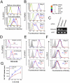
Figure 2. MS4a4B negatively regulates T cell proliferation.
A, Retrovirus-driven over-expression of MS4a4B inhibits proliferation of primary T cells. Primary CD4+ T cells isolated from spleen with anti-CD4 magnetic beads were stimulated by anti-CD3/ anti-CD28 antibodies and then were infected with MS4a4B-retroviral vector or mock vector. After infection, cells were labeled with PKH-26 Red Fluorescent linker kit. Proliferation of the tested cells was analyzed by flow cytometry (FL2). Data shown are representative of three repeat experiments. B, Forced expression of MS4a4B by LV-vector reduced proliferation of EL4 cells. Purified EL4 cells with stable lentiviral infection were labeled with PKH-26 Red and then cultured in complete RPMI 1640 medium. Cells were collected on the days indicated. Cell proliferation was assessed by flow cytometry. Data shown are representative of three repeat experiments. The numbers in histograms are mean fluorescence intensity (MFI) ± SD. ***, p<0.001. C, Knockdown of MS4a4B by siRNA. T32 cells were transfected with either FAM-labeled negative control siRNA or FAM-labeled MS4a4B-specific siRNAs (siMS4a4B1 and siMS4a4B2). Cells were collected on day 3 after transfection. Transcription of MS4a4B mRNA was analyzed by RT-PCR. D, Cell samples collected from culture on day 4 were subjected to MS4a4B protein detection by flow cytometry with anti-MS4a4B antibody. Data are presented as fluorescence intensity of FL-2 in FAM-positive population. Blue line: siMS4a4B2-transfected cells; red line: control siRNA-transfected cells. E and F, Knockdown of MS4a4B expression accelerated proliferation of T32 cells (E) and primary T cells (F). T32 cells or anti-CD3/anti-CD28-stimulated CD4+ primary T cells were labeled with PKH-26 Red and were transfected with FAM-labeled negative control siRNA or FAM-labeled siMS4a4B2. The labeled cells were stimulated with complete medium containing 20 U/ml IL-2. Cells were collected from culture at the time indicated for determination of proliferation rate by flow cytometric analysis. Data are presented as representative of three repeat experiments. Tinted grey peak: unlabeled cells; blue line: siMS4a4B2-transfected cells; red line: control siRNA-transfected cells. The numbers in histograms are MFI ± SD. *, p<0.05; **, p<0.01; ***, p<0.001. G, knockdown of MS4a4B accelerated IL-2-induced proliferation of T32 cells. T32 cells were infected with either shMS4a4B- or shLuc-lentiviral vector. Infected cells (1×10⁵) were cultured in 96 well plate for 24 hr in the presence of serially diluted IL-2, followed by incubation with 1μCi 3H-thymidine for an additional 16 hr. Cell proliferation was assayed by 3H-thymidine incorporation. A representative of two independent experiments is shown. *, p<0.05; **, p<0.01.
Figure 3
Figure 3. Expression of MS4a4B by lentiviral vector inhibits propagation of thymoma cells in vivo.
EL4 thymoma cells were infected by MS4a4B-LV or mock LV control. The infected cells (GFP+) were purified by flow cytometric cell sorting (purity: >99%). 1.5×10⁷ sorted cells were infused into C57BL/6J recipients by i.v injection. Mice were sacrificed on day 3 after cell transfer. EL4 thomoma cells (GFP+) in blood and spleen were assessed by flow cytometry. Data shown are representative of three repeat experiments. A, Representative flow histograms for each group of blood samples. The number shown is percentage of GFP+ cells in lymphocyte gate. B, Percentage ± SD of GFP+ cells in blood samples from mice injected with either MS4a4B-LV-infected or mock LV-infected EL4 cells (N = 5). C, Representative flow histograms for each group of spleen samples. The number shown is the percentage of GFP+ cells in lymphocyte gate. D, Percentage ± SD of GFP+ cells in spleens from mice injected with either MS4a4B-LV-infected or mock LV-infected EL4 cells (N = 5). E, MS4a4B inhibits solid thymoma growth in vivo. 1×10⁶ EL4 cells were injected subcutaneously in the right flank of mice. Size of tumor was measured daily after inoculation. Results are presented as mean ± SD of mm2 (N = 8). Data shown are representative of two independent repeat experiments.
Figure 4
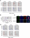
Figure 4. Expression of MS4a4B in T cells modulates cell cycle progression.
A, MS4a4B-LV or mock LV-infected EL4 thymoma cells were synchronized by treatments with 2.5 mM thymidine and serum starvation. The synchronized cells were adjusted into 5 × 10⁵/ml and were stimulated with complete medium containing 15% FCS. For cell cycle analysis, cells were harvested from culture at the time indicated. DNA content in cells was determined by propidium iodide staining and flow cytometric analysis. Results are shown as histogram from each sample with numbers indicating percentage of S-G2/M phase cells. Data presented are representative of three repeat experiments. B, Infection of T32 cells by shMS4a4B-expressing LV vector decreased MS4a4B protein expression. T32 cells stably infected with shMS4a4B-LV or shLuc-LV control were sorted by flow cytometry. MS4a4B expression in the sorted T32 cells was assessed by flow cytometry with anti-MS4a4B staining. Data are shown as representative histograms from analysis with Flowjo. C, shMS4a4B-LV or shLuc-LV control-infected T32 were co-stained with DAPI and anti-MS4a4B antibody, followed by labeling with anti-rabbit IgG-Cy3 conjugate. Knockdown of MS4a4B protein in T32 cells by shMS4a4B-LV was examined by confocal microscopy (Magnification, ×40). D, Knockdown of MS4a4B expression in T32 cells by shMS4a4B-LV inhibited cell cycle progression. shMS4a4B-LV or shLuc-LV control-infected T32 cells were synchronized as described in “A”. Synchronized cells were stimulated by 15% FCS RPMI medium containing 20 U/ml IL-2. Cell samples were harvested from culture at the time indicated. Cell cycle was analyzed by propidium iodide staining. Data are shown as the representative histogram of three repeat experiments. The numbers in histogram are the percentage of cells in S-G2/M phase.
Figure 5
Figure 5. Expression of cell cycle-regulatory genes in MS4a4B-LV-infected EL4 cells.
A, Total RNA was isolated from MS4a4B-LV vector- or mock LV vector-infected EL4 cells. Expression of 84 key regulatory genes on cell cycle progression was determined with real time PCR array analysis kit (Cat#: PAMM-020, SABiosciences, Gaithersburg, MD). Data were analyzed with on-line analysis software according to the manufacturer's instructions and presented as scatter plot with fold change of genes. Red dot: genes increased by ≥4 fold; blue dot: genes decreased by ≥4 fold. B, Genes with fold change ≥4 were shown as column figure. C, MS4a4B (or control) vector-infected EL4 cells were synchronized and stimulated with 15% FCS for 8 hours. Cell lysates (100 μg) were immunoprecipitated with anti-Cdk2-protein A beads. Cdk2-cyclin activity was assayed as described in “Methods”. The same samples (25 μg) were used for western blot with anti-β-actin antibody to ensure identical loading. Relative intensity (sample vs. LV control) is shown in the lower panel. *, p<0.05. D, Cell lysates described in “C” were separated on 10% SDS gel followed by western blotting with appropriate antibodies as indicated. E. Relative intensity of blots in “D” (sample vs. internal control (β-actin)) was determined by densitometry. *, p<0.05.

References (VSports手机版)

    1. Xu H, Williams MS, Spain LM. Patterns of expression, membrane localization, and effects of ectopic expression suggest a function for MS4a4B, a CD20 homolog in Th1 T cells. Blood. 2006;107:2400–2408. - PMC - PubMed
    1. Ishibashi K, Suzuki M, Sasaki S, Imai M. Identification of a new multigene four-transmembrane family (MS4A) related to CD20, HTm4 and β subunit of the high-affinity IgE receptor. Gene. 2001;264:87–93. - PubMed
    1. Liang Y, Tedder TF. Identification of a CD20-, FcεRIβ-, and HTm4-related gene family: sixteen new MS4A family members expressed in human and mouse. Genomics. 2001;72:119–127. - PubMed
    1. Liang Y, Buckley TR, Tu L, Langdon SD, Tedder TF. Structural organization of the human MS4A gene cluster on Chromosome 11q12. Immunogenetics. 2001;53:357–368. - PubMed
    1. Tedder TF, Disteche CM, Louie E, Adler DA, Croce CM, et al. The gene that encodes the human CD20 (B1) differentiation antigen is located on chromosome 11 near the t(11;14)(q13;q32) translocation site. J Immunol. 1989;142:2555–2559. - PubMed

Publication types

MeSH terms

Substances (V体育官网入口)