Skip to main page content
U.S. flag

An official website of the United States government

Dot gov

The . gov means it’s official. Federal government websites often end in . gov or . mil. Before sharing sensitive information, make sure you’re on a federal government site. VSports app下载.

Https

The site is secure. The https:// ensures that you are connecting to the official website and that any information you provide is encrypted and transmitted securely V体育官网. .

. 2010 Nov;137(22):3899-910.
doi: 10.1242/dev.050021.

Macrophages define dermal lymphatic vessel calibre during development by regulating lymphatic endothelial cell proliferation

Affiliations

Macrophages define dermal lymphatic vessel calibre during development by regulating lymphatic endothelial cell proliferation

VSports最新版本 - Emma J Gordon et al. Development. 2010 Nov.

Erratum in

  • Development. 2011 Feb;138(4):797

Abstract

Macrophages have been suggested to stimulate neo-lymphangiogenesis in settings of inflammation via two potential mechanisms: (1) acting as a source of lymphatic endothelial progenitor cells via the ability to transdifferentiate into lymphatic endothelial cells and be incorporated into growing lymphatic vessels; and (2) providing a crucial source of pro-lymphangiogenic growth factors and proteases. We set out to establish whether cells of the myeloid lineage are important for development of the lymphatic vasculature through either of these mechanisms. Here, we provide lineage tracing evidence to demonstrate that lymphatic endothelial cells arise independently of the myeloid lineage during both embryogenesis and tumour-stimulated lymphangiogenesis in the mouse, thus excluding macrophages as a source of lymphatic endothelial progenitor cells in these settings. In addition, we demonstrate that the dermal lymphatic vasculature of PU. 1(-/-) and Csf1r(-/-) macrophage-deficient mouse embryos is hyperplastic owing to elevated lymphatic endothelial cell proliferation, suggesting that cells of the myeloid lineage provide signals that act to restrain lymphatic vessel calibre in the skin during development VSports手机版. In contrast to what has been demonstrated in settings of inflammation, macrophages do not comprise the principal source of pro-lymphangiogenic growth factors, including VEGFC and VEGFD, in the embryonic dermal microenvironment, illustrating that the sources of patterning and proliferative signals driving embryonic and disease-stimulated lymphangiogenesis are likely to be distinct. .


V体育官网入口 - Figures

Fig. 1.
Fig. 1.
LYVE1-positive macrophages are intimately associated with lymphatic vessels but do not express PROX1. Confocal z-stack images of 60 μm sections demonstrating the localisation of LYVE1-positive, GFP-positive macrophages in close association with, and apparently integrated within (arrowheads), the jugular lymph sac (JLS; A-C) and dermal lymphatic vessel (DLV; D-G) endothelium of E14.5 c-fmsEGFP embryos. G illustrates a 90° rotation of F in order to view the lymphatic vessel in cross-section. Blue and white arrowheads denote two macrophages that appear integrated within this DLV, both of which express GFP, LYVE1 and F4/80. LYVE1-positive macrophages that are apparently integrated within the JLS or DLV endothelium do not express PROX1 (H-O). White circles in K and O illustrate the location of macrophages (arrows) in J and N. K and O illustrate single-channel PROX1 images of J and N, respectively. Data are representative of at least four independent experiments. Scale bars: 150 μm in A; 100 μm in D,H,L; 50 μm in B,E,I,M; 25 μm in C,F,J,K,N,O.
Fig. 2.
Fig. 2.
Lineage tracing in Csf1r-iCre;Z/EG embryos reveals that cells of the myeloid lineage do not comprise a pool of lymphatic endothelial progenitor cells during development. Confocal z-stack images of 60 μm sections demonstrating the localisation of LYVE1-positive, EGFP-positive macrophages in close association with, and apparently integrated within (arrows), the JLS (A-C) and DLV (D-F) endothelium of E14.5 Csf1r-iCre;Z/EG embryos. LYVE1-positive macrophages within the JLS or DLV endothelium do not express PROX1 (C,F). White circles illustrate the location of macrophages (arrows) in B and E. C and F illustrate single-channel PROX1 images of B and E, respectively. Data are representative of at least three independent experiments. Scale bars: 100 μm in A,D; 25 μm in B,C,E,F.
Fig. 3.
Fig. 3.
Myeloid cells do not comprise a pool of lymphatic endothelial progenitors during tumour-stimulated lymphangiogenesis. Lineage tracing in adult LysMCre+/–;ROSA26R+/– mice revealed select β-galactosidase-positive cells derived from the myeloid lineage apparently integrated within PROX1-positive, CD31-positive pre-existing dermal (A-D) or newly generated peri- or intra-tumoural (E-H) lymphatic vessels. PROX1 expression was not observed in any β-galactosidase-positive cells within lymphatic vessels (C,G, arrows). White circles in D and H illustrate the location of macrophages (arrows) in C and G, respectively. D and H illustrate single-channel PROX1 images of C and G, respectively. Data are representative of eight independent experiments per tumour type. Illustrated is an example of the LLC tumour. Scale bars: 100 μm in A,E; 50 μm in B,F; 25 μm in C,D,G,H.
Fig. 4.
Fig. 4.
Dermal lymphatic vessel hyperplasia in macrophage-deficient mice. Whole-mount immunostaining and confocal microscopy of E16.5 dorsal skin from mice of the indicated genotype revealed that PROX1- (A-C) and NRP2- (D-F) positive dermal lymphatic vessels are hyperplastic in macrophage-deficient PU.1–/– (B,E) and Csf1r–/– (F) embryos compared with wild-type (A,D) and Rag2–/– (C) counterparts. The absence of macrophages in PU.1–/– embryos was confirmed by F4/80 immunostaining (B). Vessel hyperplasia is restricted to the lymphatic vasculature; patterning and calibre of CD31-positive blood vessels (D-F) in embryonic PU.1–/– (E) and Csf1r–/– (F) skin is indistinguishable from that of wild type (D). Vessel diameter was quantified using LVAP and ImageJ software (G). Data are representative of at least six independent experiments, with the exception of Csf1r–/– analysis, where n=3. Data shown represent the mean ± s.e.m. P-values were calculated using Student's paired t-test. *, P≤0.05. Scale bars: 150 μm in A-F.
Fig. 5.
Fig. 5.
Lymphatic vessel branching and proliferation in wild-type and PU.1–/– embryos. Whole-mount staining of dorsal skin isolated from E16.5 wild-type and PU.1–/– embryos stained with antibodies against NRP2 and PH3. White dots denote branch-points (A,B). Arrows illustrate PH3-positive, NRP2-positive mitotic lymphatic endothelial cells (D,E). At least four independent embryos of each genotype were analysed, at least three (for branching analysis, C) or six (for proliferation analysis, F) fields of view were quantified per embryo. Data shown represent the mean ± s.e.m. P-values were calculated using Student's paired t-test. *, P≤0.05. Scale bars: 150 μm in A,B; 100 μm in D,E.
Fig. 6.
Fig. 6.
Angiopoietin 2 is a pro-survival signal in lymphatic endothelial cells. (A-D) Whole-mount immunostaining and confocal microscopy of dorsal skin from an allelic series of E16.5 wild-type (A), PU.1–/–;Ang2+/+ (B), PU.1–/–;Ang2+/– (C) and PU.1+/+;Ang2–/– (D) embryos demonstrates rescue of lymphatic vessel hyperplasia in PU.1–/–;Ang2+/– skin (C compared with B) and pronounced lymphatic vessel hypoplasia in PU.1+/+;Ang2–/– skin (D). The patterning and recruitment of vascular smooth muscle to the large arteries and veins in the dorsal skin of mutant mice (B-D) is indistinguishable from that of wild-type littermates (A). NRP2-positive hair follicles are particularly pronounced in Ang2–/– skin (D). (E) Lymphatic vessel diameter was quantified using LVAP and ImageJ software. Data are representative of at least four independent experiments. Data shown represent the mean ± s.e.m. P-values were calculated using Student's paired t-test. *, P≤0.05. (F,G) Whole-mount immunostaining and confocal microscopy of wild-type (F) and PU.1–/– (G) dorsal skin stained with antibodies to activated caspase-3 and VEGFR3. (H-J) Immunostaining of transverse sections through c-fmsEGFP jugular lymph sacs stained with LYVE1 and activated caspase-3 antibodies. J is a single-channel image of I illustrating a caspase-3-positive apoptotic cell (arrowhead). Scale bars: 150 μm in A-D; 100 μm in F,G; 100 μm in H; 25 μm in I,J.
Fig. 7.
Fig. 7.
PU.1–/– embryos exhibit hypoplastic jugular lymph sacs. (A-F) Immunostaining of transverse sections through the jugular region of E14.5 wild-type (A-C) and PU.1–/– (D-F) embryos with antibodies to PROX1 and CD31. Dashed white lines outline the margins of jugular lymph sacs at each level analysed (L1-L3, indicated at left). (G) JLS size was quantified using ImageJ software. At least four independent embryos of each genotype were analysed. Data shown represent the mean ± s.e.m. P-values were calculated using Student's paired t-test. *, P≤0.05. Scale bars: 100 μm in A-F.
Fig. 8.
Fig. 8.
Expression of pro-lymphangiogenic factors is elevated in the skin of PU.1–/– mice. Semi-quantitative RT-PCR was used to determine the relative level of gene expression in PU.1–/– (n=4) compared with wild-type (n=4) E16.5 skin samples. Data are depicted as the percentage change in mRNA expression in PU.1–/– compared with wild-type skin and represent the mean ± s.e.m. No change in expression between PU.1–/– and wild-type skin is represented by 0%. P-values were calculated using Student's paired t-test. *, P≤0.05.
Fig. 9.
Fig. 9.
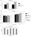
Regulation of lymphatic endothelial cell proliferation in vitro. (A,B) Co-culture of primary embryonic dermal macrophages (A), but not macrophage or skin-cell-conditioned media (B), with primary embryonic dermal lymphatic endothelial cells resulted in elevated lymphatic endothelial cell proliferation. Addition of VEGFR3/Fc to conditioned media assays did not inhibit lymphatic endothelial cell proliferation. (C) siRNA-mediated knockdown of Tie1 in primary embryonic dermal lymphatic endothelial cells resulted in significantly decreased lymphatic endothelial cell proliferation. Data shown represent the mean ± s.e.m. of three independent experiments. P-values were calculated using Student's paired t-test. *, P≤0.05.

References

    1. Abramoff M. D., Magelhaes M. P., Ram S. J. (2004). Image processing with ImageJ. Biophotonics Int. 11, 36-42
    1. Alitalo K., Tammela T., Petrova T. V. (2005). Lymphangiogenesis in development and human disease. Nature 438, 946-953 - VSports注册入口 - PubMed
    1. Back J., Dierich A., Bronn C., Kastner P., Chan S. (2004). PU.1 determines the self-renewal capacity of erythroid progenitor cells. Blood 103, 3615-3623 - VSports在线直播 - PubMed
    1. Baluk P., Tammela T., Ator E., Lyubynska N., Achen M. G., Hicklin D. J., Jeltsch M., Petrova T. V., Pytowski B., Stacker S. A., et al. (2005). Pathogenesis of persistent lymphatic vessel hyperplasia in chronic airway inflammation. J. Clin. Invest. 115, 247-257 - PMC - PubMed
    1. Banerji S., Ni J., Wang S. X., Clasper S., Su J., Tammi R., Jones M., Jackson D. G. (1999). LYVE1, a new homologue of the CD44 glycoprotein, is a lymph-specific receptor for hyaluronan. J. Cell Biol. 144, 789-801 - PMC - PubMed

Publication types

"VSports" MeSH terms