Skip to main page content
U.S. flag

An official website of the United States government

Dot gov

The . gov means it’s official. Federal government websites often end in . gov or VSports app下载. mil. Before sharing sensitive information, make sure you’re on a federal government site. .

Https

The site is secure. The https:// ensures that you are connecting to the official website and that any information you provide is encrypted and transmitted securely V体育官网. .

Review
. 2011 Jan;68(2):219-33.
doi: 10.1007/s00018-010-0530-4. Epub 2010 Oct 8.

Mitochondrial DNA replication and disease: insights from DNA polymerase γ mutations

Affiliations
Review

Mitochondrial DNA replication and disease: insights from DNA polymerase γ mutations

Jeffrey D Stumpf et al. Cell Mol Life Sci. 2011 Jan.

Abstract

DNA polymerase γ (pol γ), encoded by POLG, is responsible for replicating human mitochondrial DNA. About 150 mutations in the human POLG have been identified in patients with mitochondrial diseases such as Alpers syndrome, progressive external ophthalmoplegia, and ataxia-neuropathy syndromes VSports手机版. Because many of the mutations are described in single citations with no genotypic family history, it is important to ascertain which mutations cause or contribute to mitochondrial disease. The vast majority of data about POLG mutations has been generated from biochemical characterizations of recombinant pol γ. However, recently, the study of mitochondrial dysfunction in Saccharomyces cerevisiae and mouse models provides important in vivo evidence for the role of POLG mutations in disease. Also, the published 3D-structure of the human pol γ assists in explaining some of the biochemical and genetic properties of the mutants. This review summarizes the current evidence that identifies and explains disease-causing POLG mutations. .


Figures
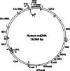
Fig. 1
Fig. 1
Schematic of the mitochondrial genome. Single letters represent the amino acid code for the specific mitochondrial tRNA. The H strand is replicated clockwise beginning at OriH. The L strand is replicated counter-clockwise beginning at OriL
Fig. 2
Fig. 2
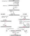
Schematic of the base excision repair pathways present in mitochondria. The damaged base is excised by a glycosylase to create an abasic site. APE1 endonuclease generates a strand break leaving a 5′ deoxyribose phosphate (5′ dRP) downstream of the lesion. Pol γ participates in either the single-nucleotide (SN-BER) or the long patch (LP-BER) base excision repair pathway. In SN-BER, Pol γ fills the nucleotide gap and removes the 5′ dRP, allowing for ligation by Ligase III. In LP-BER, Pol γ performs strand-displacement synthesis producing a 5′ flap that is excised by DNA2 and FEN1. The resulting substrate is ligated by Ligase III
Fig. 3
Fig. 3
Schematic diagram of human pol γ gene (top) and protein (bottom) showing the location of amino acid substitutions resulting from mutations associated with disease (in the boxes) and neutral polymorphisms found in unaffected populations (arrows). The top line displays the relative positions of the exons. The gene is separated into the mitochondrial targeting sequence (green), exonuclease domain (brown), linker domain, and the polymerase domain (blue)
Fig. 4
Fig. 4
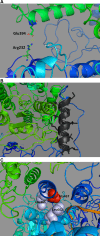
The 3-dimensional structure of the human DNA polymerase γ holoenzyme depicting several residues involved in disease and important for interaction between the catalytic pol γ-α subunit (blue) and the accessory pol γ-β subunit (green). These illustrations were derived using PDB 3IKM [74] in the program PyMOL (http://www.pymol.org/). a The salt bridge between Arg232 of the pol gamma catalytic subunit and Glu394 of the distal p55 accessory subunit. b Pol γ- α amino acids 543–558 (shown in black) form a helix of hydrophobic residues that stabilize interactions with the proximal pol γ- β subunit. c Ala467 is located near Leu466 and Leu602, which is postulated to comprise an important hydrophobic environment for subunit interaction
Fig. 5
Fig. 5
The 3-dimensional structure of the human DNA polymerase γ holoenzyme depicting several residues involved in disease. These illustrations were derived using PDB 3IKM [74] in the program PyMOL (http://www.pymol.org/). a Active site of the human DNA polymerase gamma illuminating residues 848, 851, 852, and 853 that are altered in Alpers patients. The side chain of Arg853 is in a position to provide electrostatic interaction with Asp1135, which along with Glu1136, chelate the two active site Mg2+ ions. b The side chain of Arg807 is shown here to protrude into the DNA binding cleft in the open structure. Although the polymerase is in the ‘closed’ conformation, DNA is modeled into its predicted location in an ‘open’ conformation

VSports手机版 - References

    1. Bebenek K, Kunkel TA. Functions of DNA polymerases. Adv Protein Chem. 2004;69:137–165. - PubMed
    1. Ropp PA, Copeland WC. Cloning and characterization of the human mitochondrial DNA polymerase, DNA polymerase gamma. Genomics. 1996;36:449–458. - PubMed
    1. Sweasy JB, Lauper JM, Eckert KA. DNA polymerases and human diseases. Radiat Res. 2006;166:693–714. - VSports - PubMed
    1. Graziewicz MA, Longley MJ, Copeland WC. DNA polymerase gamma in mitochondrial DNA replication and repair. Chem Rev. 2006;106:383–405. - VSports手机版 - PubMed
    1. Lim SE, Longley MJ, Copeland WC. The mitochondrial p55 accessory subunit of human DNA polymerase gamma enhances DNA binding, promotes processive DNA synthesis, and confers N-ethylmaleimide resistance. J Biol Chem. 1999;274:38197–38203. - PubMed

V体育官网 - Publication types

MeSH terms

"V体育安卓版" Substances