Skip to main page content
U.S. flag

An official website of the United States government

Dot gov

The . gov means it’s official. Federal government websites often end in . gov or VSports app下载. mil. Before sharing sensitive information, make sure you’re on a federal government site. .

Https

The site is secure. The https:// ensures that you are connecting to the official website and that any information you provide is encrypted and transmitted securely V体育官网. .

. 2010 Oct 12;107(41):17686-91.
doi: 10.1073/pnas.1012016107. Epub 2010 Sep 22.

Induction of regulatory T cells by macrophages is dependent on production of reactive oxygen species

Affiliations

V体育2025版 - Induction of regulatory T cells by macrophages is dependent on production of reactive oxygen species

Marina D Kraaij et al. Proc Natl Acad Sci U S A. .

"V体育安卓版" Abstract

The phagocyte NAPDH-oxidase complex consists of several phagocyte oxidase (phox) proteins, generating reactive oxygen species (ROS) upon activation VSports手机版. ROS are involved in the defense against microorganisms and also in immune regulation. Defective ROS formation leads to chronic granulomatous disease (CGD) with increased incidence of autoimmunity and disturbed resolution of inflammation. Because regulatory T cells (Tregs) suppress autoimmune T-cell responses and are crucial in down-regulating immune responses, we hypothesized that ROS deficiency may lead to decreased Treg induction. Previously, we showed that in p47(phox)-mutated mice, reconstitution of macrophages (Mph) with ROS-producing capacity was sufficient to protect the mice from arthritis. Now, we present evidence that Mph-derived ROS induce Tregs. In vitro, we showed that Mph ROS-dependently induce Treg, using an NADPH-oxidase inhibitor. This finding was confirmed genetically: rat or human CGD Mph with mutated p47(phox) or gp91(phox) displayed hampered Treg induction and T-cell suppression. However, basal Treg numbers in these subjects were comparable to those in controls, indicating a role for ROS in induction of peripheral Tregs. Induction of allogeneic delayed-type hypersensitivity with p47(phox)-mutated Mph confirmed the importance of Mph-derived ROS in Treg induction in vivo. We conclude that NAPDH oxidase activity in Mph is important for the induction of Tregs to regulate T cell-mediated inflammation. .


Conflict of interest statement

The authors declare no conflict of interest.

Figures

Fig. 1.
Fig. 1.
Mph produce ROS. (A) mRNA and (B) protein expression levels of the two most important components of the Nox2 complex, p47phox and gp91phox, determined in DC and Mph by RT-PCR and flow cytometry, respectively. In A mRNA expression levels were corrected for GAPDH expression, and expression levels of DC were set to 1. B shows the ratio between specific staining and isotype control. Results shown are average and SD of the relative expression levels of four experiments with cells from four different donors. (C) The capacity of DC and Mph to produce ROS was measured after PMA stimulation. Results shown are average and SD of 6–10 independent experiments; the conditions after simulation with PMA or with vehicle (DMSO) are shown. (D) Representative FACS histograms of ROS production, measured by DHR123 fluorescence, by DC (Left) and Mph (Right) after PMA (dotted line) or DMSO (control; solid line) stimulation. (E) Production of ROS by DC and Mph was measured after PMA or vehicle (DMSO) activation and in the absence or presence of the specific p47phox inhibitor apocynin (1 mM). Results shown are the average and SD of three or four independent experiments. *P < 0.05.
Fig. 2.
Fig. 2.
Mph suppress T-cell activation in a ROS-dependent fashion. T cells (150,000) were activated with anti-CD3/28 Ab, and Mph or DC were added in increasing numbers (x axes). After 5 d of coculture, IFN-γ production (A) and proliferation (D) were determined by ELISA and 3H thymidine incorporation, respectively. Results shown are the average and SD of a representative experiment performed in triplicate. (B and E) We cocultured 150,000 T cells and 25,000 Mph in the absence or presence of different concentrations of apocynin (x axis) to study the effect of ROS on Mph-mediated T-cell suppression. After 5 d of coculture, IFN-γ production (B) and proliferation (E) were determined by ELISA and 3H thymidine incorporation, respectively. Representative experiments are shown. (C and F) Experiments were similar to those in A, B, D, and E, but the average and SD of three or four independent experiments are shown for an APC:T cell ratio of 1:6. Values shown are relative to the conditions without APC (100%, white bars). *P < 0.05
Fig. 3.
Fig. 3.
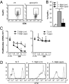
Mph induce Tregs via ROS. (A) Expression of FoxP3 within the CD3+CD4+CD25+ population of T cells primed with Mph in the absence (Left, ctr) or presence (Right) of apocynin. (B) CD4+CD25 T cells were primed with Mph in the presence [T + Mph (apo)] or absence [T + Mph (ctr)] of apocynin for 7 d, and then the percentage of FoxP3+ cells among CD3+CD4+CD25+ cells was analyzed. The white bar (T) represents CD4+CD25 T cells (cultured similarly but without Mph. Results shown are the average and SD of three experiments. (C) T cells were primed with (Left) or without (Right) Mph in presence or absence of apocynin (1 mM). After 5 d, these T cells were used as suppressor cells (S) and were combined with responder T cells (R) and irradiated feeder cells (F). Proliferation of responder cells was assessed by 3H thymidine incorporation (C) or by determining CFSE dilution (D). C shows average and SD of four experiments. In D, the 1:1 ratio from a representative experiment of three is shown, as well as the control condition without S but with twice the number of responders (F2R) to correct for crowding effects. *P < 0.05.
Fig. 4.
Fig. 4.
CGD Mph induce fewer Tregs. (A) The percentage of FoxP3+ cells among CD4+CD25+ cells in peripheral blood of CGD patients and healthy controls (ctr). (B) ROS production by Mph differentiated from healthy controls or CGD monocytes, as determined by flow cytometry after DHR123 staining. The ratio of ROS production after stimulation with PMA or DMSO is depicted. (C) CD4+CD25 T cells (106 per condition) from a single allogeneic donor were primed with Mph from either CGD patients or controls in presence of anti-CD3/28 Ab. After 5 d the number of viable T cells was determined. (D) Representative T-cell clustering observed after activation with anti-CD3/CD28 in presence of control Mph (Upper) or Mph derived from CGD patients (Lower). (Scale bars: 25 μm.) (E) T cells primed with Mph derived from CGD patients or controls were analyzed by flow cytometry for the percentage of FoxP3+ cells among CD4+CD25+ cells. (F) CFSE-labeled responder cells were cocultured with the T cells that were primed with ctr or CGD Mph and irradiated feeder cells (F2R) in the presence of anti-CD3/28, and dilution of CFSE was measured by flow cytometry after 4 d. (G) In parallel experiments, 3H thymidine incorporation was determined. Gray dots are control conditions in the absence of Mph-primed suppressor T cells but with double amounts of responder cells (F2R) to correct for crowding effects. In the supernatants of this suppression assay, IFN-γ (H) and IL-17 (I) levels were determined. *P < 0.05.
Fig. 5.
Fig. 5.
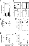
Mph suppress DTH responses in vivo in a ROS-dependent fashion. (A) Rat bone marrow cells were cultured with rat GM-CSF and IL-4 or human M-CSF to obtain DC and Mph, respectively. ROS production by PMA-stimulated DC and Mph generated from DA.Ncf1DA/DA or congenic Ncf1-wild-type DA.Ncf1E3/E3 rats was measured by DHR123 staining. Results shown are average and SD of four experiments. (B) Treg gating strategy on peripheral blood. (C) The percentage of FoxP3+ cells among CD3+CD4+CD25+ cells in peripheral blood of naïve DA.Ncf1E3/E3 (E3/E3) rats or DA.Ncf1DA/DA (DA/DA) rats. (D) Lewis rats were primed with Mph from DA.Ncf1DA/DA rats (DA/DA) or DA.Ncf1E3/E3 rats (E3/E3) or with PBS at day 0. At day 11, the percentage of FoxP3+ cells among CD3+CD4+CD25+ T cells was determined and compared with the levels in naïve Lewis rats. (E) After rats were immunized at day 11 and challenged in the ear at day 25 with irradiated DA/DA splenocytes, the difference in ear thickness, as a measure for the DTH reaction, was determined at day 26. (F) The percentage of FoxP3+ cells among CD3+CD4+CD25+ T cells at day 26 after priming. *P < 0.05.

References

    1. Hultqvist M, Olsson LM, Gelderman KA, Holmdahl R. The protective role of ROS in autoimmune disease. Trends Immunol. 2009;30:201–208. - PubMed
    1. Ghezzi P, Bonetto V, Fratelli M. Thiol-disulfide balance: From the concept of oxidative stress to that of redox regulation. Antioxid Redox Signal. 2005;7:964–972. - "V体育2025版" PubMed
    1. Gelderman KA, et al. Rheumatoid arthritis: The role of reactive oxygen species in disease development and therapeutic strategies. Antioxid Redox Signal. 2007;9:1541–1567. - PubMed
    1. Kazama H, et al. Induction of immunological tolerance by apoptotic cells requires caspase-dependent oxidation of high-mobility group box-1 protein. Immunity. 2008;29:21–32. - PMC - PubMed
    1. Corzo CA, et al. Mechanism regulating reactive oxygen species in tumor-induced myeloid-derived suppressor cells. J Immunol. 2009;182:5693–5701. - PMC - PubMed

Publication types (VSports手机版)

VSports最新版本 - MeSH terms