Nonlinear time course of brain volume loss in cognitively normal and impaired elders
- PMID: 20855131
- PMCID: PMC3032014 (V体育官网)
- DOI: 10.1016/j.neurobiolaging.2010.07.012 (VSports app下载)
V体育官网 - Nonlinear time course of brain volume loss in cognitively normal and impaired elders
Abstract
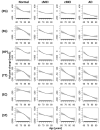
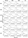
The goal was to elucidate the time course of regional brain atrophy rates relative to age in cognitively normal (CN) aging, mild cognitively impairment (MCI), and Alzheimer's disease (AD), without a priori models for atrophy progression. Regional brain volumes from 147 cognitively normal subjects, 164 stable MCI, 93 MCI-to-AD converters and 111 ad patients, between 51 and 91 years old and who had repeated 1. 5 T magnetic resonance imaging (MRI) scans over 30 months, were analyzed. Relations between regional brain volume change and age were determined using generalized additive models, an established nonparametric concept for approximating nonlinear relations. Brain atrophy rates varied nonlinearly with age, predominantly in regions of the temporal lobe. Moreover, the atrophy rates of some regions leveled off with increasing age in control and stable MCI subjects whereas those rates progressed further in MCI-to-AD converters and AD patients VSports手机版. The approach has potential uses for early detection of AD and differentiation between stable and progressing MCI. .
Copyright © 2012 Elsevier Inc. All rights reserved. V体育安卓版.
VSports app下载 - Conflict of interest statement
The remaining authors have no potential financial or personal conflicts of interest including relationships with other people or organizations within 3 years of beginning the work submitted that could inappropriately influence their work.
Figures


V体育官网入口 - References
-
- Apostolova LG, Hwang KS, Andrawis JP, Green AE, Babakchanian S, Morra JH, Cummings JL, Toga AW, Trojanowski JQ, Shaw LM, Jack CR, Jr, Petersen RC, Aisen PS, Jagust WJ, Koeppe RA, Mathis CA, Weiner MW, Thompson PM. 3D PIB and CSF biomarker associations with hippocampal atrophy in ADNI subjects. Neurobiol Aging. S0197-4580(10)00214-9. - PMC - PubMed
-
- Ashburner J, Friston KJ. Voxel-based morphometry--the methods. Neuroimage. 2000;11(6 Pt 1):805–21. - PubMed
-
- Braak H, Braak E. Evolution of the neuropathology of Alzheimer’s disease. Acta Neurol Scand Suppl. 1996;165:3–12. - PubMed
-
- Chen JH, Lin KP, Chen YC. Risk factors for dementia. J Formos Med Assoc. 2009;108(10):754–64. - PubMed
-
- Davatzikos C, Xu F, An Y, Fan Y, Resnick SM. Longitudinal progression of Alzheimer’s-like patterns of atrophy in normal older adults: the SPARE-AD index. Brain. 2009;132(Pt 8):2026–35. - VSports在线直播 - PMC - PubMed
Publication types
- "VSports注册入口" Actions
- VSports app下载 - Actions
MeSH terms
- "VSports最新版本" Actions
- V体育官网 - Actions
- "V体育官网入口" Actions
- Actions (VSports app下载)
- Actions (V体育安卓版)
Grants and funding
- K01 AG030514/AG/NIA NIH HHS/United States
- P50 AG023501/AG/NIA NIH HHS/United States
- R01 NS031966/NS/NINDS NIH HHS/United States (VSports手机版)
- R01 AG010897/AG/NIA NIH HHS/United States
- VSports - U19 AG024904/AG/NIA NIH HHS/United States
- V体育安卓版 - T32 EB001631-05/EB/NIBIB NIH HHS/United States
- P01 AG019724/AG/NIA NIH HHS/United States
- P01 AG012435/AG/NIA NIH HHS/United States
- VSports最新版本 - P41 RR023953/RR/NCRR NIH HHS/United States
- U24 RR021992/RR/NCRR NIH HHS/United States
- T32 EB001631/EB/NIBIB NIH HHS/United States
- P30 AG010129/AG/NIA NIH HHS/United States
- U01 AG024904/AG/NIA NIH HHS/United States
- U19 AG010483/AG/NIA NIH HHS/United States
LinkOut - more resources
Full Text Sources
Medical

