Skip to main page content
U.S. flag

An official website of the United States government

Dot gov

The . gov means it’s official. Federal government websites often end in . gov or . mil VSports app下载. Before sharing sensitive information, make sure you’re on a federal government site. .

Https

The site is secure. The https:// ensures that you are connecting to the official website and that any information you provide is encrypted and transmitted securely V体育官网. .

. 2010 Sep 14;18(3):231-43.
doi: 10.1016/j.ccr.2010.08.007.

An ARF-independent c-MYC-activated tumor suppression pathway mediated by ribosomal protein-Mdm2 Interaction (VSports注册入口)

Affiliations

VSports最新版本 - An ARF-independent c-MYC-activated tumor suppression pathway mediated by ribosomal protein-Mdm2 Interaction

Everardo Macias et al. Cancer Cell. .

Abstract (VSports在线直播)

In vitro studies have shown that inhibition of ribosomal biogenesis can activate p53 through ribosomal protein (RP)-mediated suppression of Mdm2 E3 ligase activity. To study the physiological significance of the RP-Mdm2 interaction, we generated mice carrying a cancer-associated cysteine-to-phenylalanine substitution in the zinc finger of Mdm2 that disrupted its binding to RPL5 and RPL11. Mice harboring this mutation, retain normal p53 response to DNA damage, but lack of p53 response to perturbations in ribosome biogenesis. Loss of RP-Mdm2 interaction significantly accelerates Eμ-Myc-induced lymphomagenesis. Furthermore, ribosomal perturbation-induced p53 response does not require tumor suppressor p19ARF. Collectively, our findings establish RP-Mdm2 interaction as a genuine p53 stress-signaling pathway activated by aberrant ribosome biogenesis and essential for safeguarding against oncogenic c-MYC-induced tumorigenesis VSports手机版. .


Figures (VSports手机版)

Figure 1
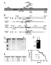
Figure 1. Generation and analysis of Mdm2C305F knockin mice
(A) A diagram of Mdm2 protein and approximate binding domains for p53, ARF, and several ribosomal proteins. (B) Schematic representation of Mdm2C305F targeting strategy. Exons 7-12 of Mdm2 are shown in black boxes. The targeting vector contains negative selection marker thymidine kinase (TK) and positive selection marker neomycin resistant gene (neo). Two loxP sites are shown as triangles. (C) Southern blot analysis was used to screen ES clones for the targeted allele. BamH1 digestion generates a 9.5 kb fragment from the wild type allele. The targeted allele gives rise to two BamH1 fragments of 4.8 kb and 6.5 kb, respectively. (D) DNA from ES cells positive for the recombined allele identified was analyzed for the presence of Mdm2C305F mutation by PCR amplification and Spe1 digestion. (E) Shown are expected and observed birth ratios from a total of 151 mice obtained from Mdm2+/m intercross. (F) Kaplan-Mierer survival curve for Mdm2+/+ (+/+), Mdm2+/C305F (+/m) and Mdm2C305F/C305F (m/m) mice are shown.
Figure 2
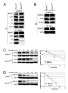
Figure 2. Characterization of Mdm2C305F mutant protein
(A) Mdm2+/+ and Mdm2m/m MEFs were treated with 5 nM Act D and lysates were immunoprecipitated (IP) with anti-Mdm2 antibody and immunoblotted (IB) for Mdm2, p53, L5 and L11 as indicated. Loading represents 5% of total lysate used for IP. (B) MEFs were treated as in (A) and lysates were analyzed as indicated. (C) Half-life of p53 in Mdm2+/+ and Mdm2m/m MEFs. Half-life assay was carried out using early passage (P1) MEFs treated with cycloheximide (50 μg/ml) and harvested with SDS lysis buffer at the indicated time points. Relative amount of p53 was quantified by densitometry, normalized to Actin and plotted. (D) Half-life of Mdm2 and Mdm2C305F. MEFs were treated and analyzed as in (C) for Mdm2.
Figure 3
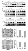
Figure 3. Mdm2C305F mice retain intact p53 response to DNA damage
(A) Mdm2+/+, Mdm2m/+ and Mdm2m/m MEFs were mock treated (Control) or treated with 1 μM doxorubicin (Dox) or 50 J/m2 of ultraviolet C (UV). The cells were harvested 18 hours post treatment and immunoblotted for p53 and Actin. (B) Mdm2+/+ and Mdm2m/m MEFs were treated with increasing dosage of Dox for 18 hrs. Cell extracts were analyzed for p53, p21 and Mdm2. (C) Induction of p21 in spleen and thymus after γ-irradiation. Spleen and thymus were harvested from Mdm2+/+ and Mdm2m/m mice 18 hours after 8-Gy whole body γ-irradiation. Protein extracts were assayed for protein levels of p53, p21, and Actin. Spleen protein extract from a p53−/− mouse was used as a negative control. (D) Apoptotic response to whole body γ-irradiation. The level of apoptosis in spleen and thymus was determined by TUNEL assay, carried out on paraffin embedded tissues from mice 18 hours after receiving 8-Gy of whole body γ-irradiation. The percentage of TUNEL positive cells was quantified and averaged from three identically treated mice. Scale bars represent 75 μm. Error bars in all cases represent ± SD.
Figure 4
Figure 4. Mdm2C305F mice demonstrate attenuated p53 response to ribosome biogenesis stress
(A) MEFs were treated with 5 nM Act D and harvested at indicated time points. Cell lysates were immunoblotted for p53 and Actin. (B) Mdm2+/+, Mdm2+/m and Mdm2m/m MEFs were either mock treated or treated with 5 nM Act D for 12 hours before harvesting for western blot analysis. (C) p53 response to a DNA damaging dosage of Act D in Mdm2m/m MEFs. MEFs were treated with 200 nM Act D for 12 hours and analyzed for p53. (D) Mouse dorsal skins were treated topically with 200 μl of acetone (Control) or Act D (1 μg/200 μl acetone). Protein lysates were prepared from snap frozen skin collected 18 hours after the treatment by epidermal scrapes and analyzed for p53. As a positive control, dorsal skins were treated with acetone and followed immediately by whole body 8-Gy γ-irradiation (IR), and the skin cells were collected 18 hours after the treatment. (E) Early passage MEFs were either mock treated or treated with 5 nM Act D for 18 hours. Cell cycle distribution was determined by flow cytometry using propidium iodide (PI) staining. Samples were run in triplicate with a minimum of 15,000 cells analyzed for each treatment. (F) BrdU incorporation analysis in Mdm2m/m keratinocytes. Mouse dorsal skins were treated as in (D) and fixed in formalin 18 hours post treatment. BrdU incorporation was determined by immunohistochemistry. For quantification, BrdU positive interfollicular basal keratinocytes from 15 representative 200 μm fields were counted. Three mice were used for each group. Asterisk indicates P value of < 0.01 determined by Tukey’s multiple comparison tests. (G) p53 response to 5-FU and MPA. MEFs were treated with 1 μM 5-FU or 2 μM MPA for 12 hours and assessed for p53 by western blotting. Error bars in all cases represent ± SD.
Figure 5
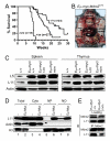
Figure 5. Myc-induced lymphomagenesis is accelerated by Mdm2C305F mutation
(A) Survival of Eμ-Myc transgenic mice. Kaplan-Mierer survival curve shows a median survival of 20, 15, and 9 weeks for Eμ-Myc;Mdm2+/+ (+/+, n= 24), Eμ-Myc;Mdm2+/m (+/m, n= 27) and Eμ-Myc;Mdm2m/m (m/m, n= 18) mice, respectively. (B) Lymphoma onset in Eμ-Myc;Mdm2m/m mice. Image shows dissection of a 58-day old Eμ-Myc;Mdm2m/m mouse with typical enlargement of liver, spleen, thymus and lymph nodes, including enlargement of Peyer’s patches and mesenteric lymph nodes. (C) Extracts from spleen and thymus of wild type (+/+) and tumor bearing Eμ-Myc transgenic mice were analyzed for L5 and L11. (D) Accumulation of L11 in nucleus in Eμ-Myc transgenic mouse splenocytes. Lysates of splenocytes from wild type and non-tumor bearing Eμ-Myc transgenic mice were subjected to fractionation as described in Experimental Procedures. The total (Total), cytoplasmic (Cyto), nucleoplasmic (NP), and nucleolar (NO) fractions were isolated and analyzed for L11. Actin (cytoplasmic) and Histone H3 (nuclear) were used as fractionation markers. (E) L11-Mdm2 binding in Eμ-Myc transgenic splenocytes. Lysates from spleens of 4-week-old, non-tumor bearing mice were immunoprecipitated with anti-Mdm2 antibody and immunoblotted as indicated. Loading represents 5% of total lysate used for IP.
Figure 6
Figure 6. p19Arf is dispensable for p53 response to nucleolar stress
(A) Oncogenic c-Myc induction of p53 in Mdm2C305F mice. Extracts from spleens of 4-week-old, non-tumor bearing Eμ-Myc;Mdm2+/+ and Eμ-Myc;Mdm2m/m transgenics as well as from their non-transgenic counterparts were analyzed for p53, p19Arf, and L11. Two mice are used in each group. (B) Myc induction of p53 in the absence of p19Arf. Early passage wild type, Mdm2m/m and p19Arf−/− MEFs were infected with retrovirus expressing either pBabe vector or pBabe-c-Myc, selected by puromycin for 3 days, then allowed to recover for 48 hours before harvesting for western analysis. (C) Early passage wild type and p19Arf−/− MEFs were treated with 5 nM Act D and harvested at the indicated time points. Cell lysates were analyzed for indicated proteins. (D) p53 response to MPA in p19Arf−/− MEFs. Early passage wild type and p19Arf−/− MEFs were treated with 2 μM MPA for 12 hours, then harvested and analyzed for p53. (E) L11-Mdm2 and p19Arf-Mdm2 interaction in Act D treated MEFs. Early passage wild type, Mdm2m/m and p19Arf−/− MEFs were treated with 5 nM Act D for 4 hours. Cell lysates were immunoprecipitated with anti-Mdm2 antibody and immunoblotted as indicated. Loading represents 5% of total lysate used for IP.
Figure 7
Figure 7. Models for RP-Mdm2-p53 signaling in response to ribosomal stress and oncogenic c-Myc
(A) In response to ribosomal stress several ribosomal proteins, including L11 and L5, bind Mdm2 and inhibit its E3 ligase function, leading to stabilization and activation of p53. (B) Oncogenic c-Myc induces high expression of both p19Arf and ribosomal proteins. The RP-Mdm2-p53 and p19Arf-Mdm2-p53 represent two parallel signaling pathways in response to oncogenic c-Myc stimulation.

References

    1. Adams JM, Harris AW, Pinkert CA, Corcoran LM, Alexander WS, Cory S, Palmiter RD, Brinster RL. The c-myc oncogene driven by immunoglobulin enhancers induces lymphoid malignancy in transgenic mice. Nature. 1985;318:533–538. - PubMed
    1. Alitalo K, Koskinen P, Makela TP, Saksela K, Sistonen L, Winqvist R. myc oncogenes: activation and amplification. Biochim Biophys Acta. 1987;907:1–32. - PubMed
    1. Allison AC. Mechanisms of action of mycophenolate mofetil. Lupus. 2005;14(Suppl 1):s2–8. - PubMed
    1. Ayrault O, Andrique L, Seite P. Involvement of the transcriptional factor E2F1 in the regulation of the rRNA promoter. Exp Cell Res. 2006;312:1185–1193. - PubMed (V体育官网入口)
    1. Barkic M, Crnomarkovic S, Grabusic K, Bogetic I, Panic L, Tamarut S, Cokaric M, Jeric I, Vidak S, Volarevic S. The p53 tumor suppressor causes congenital malformations in Rpl24-deficient mice and promotes their survival. Mol Cell Biol. 2009;29:2489–2504. - PMC - PubMed

Publication types

"VSports最新版本" MeSH terms

VSports手机版 - Substances