Skip to main page content (VSports最新版本)
U.S. flag

An official website of the United States government

Dot gov

The . gov means it’s official. Federal government websites often end in . gov or . mil VSports app下载. Before sharing sensitive information, make sure you’re on a federal government site. .

Https

The site is secure. The https:// ensures that you are connecting to the official website and that any information you provide is encrypted and transmitted securely V体育官网. .

. 2010 Sep 21;107(38):16619-24.
doi: 10.1073/pnas.1010722107. Epub 2010 Sep 7.

Notch signaling contributes to proliferation and tumor formation of human T-cell leukemia virus type 1-associated adult T-cell leukemia

Affiliations

"VSports最新版本" Notch signaling contributes to proliferation and tumor formation of human T-cell leukemia virus type 1-associated adult T-cell leukemia

Joanna Pancewicz et al. Proc Natl Acad Sci U S A. .

Abstract

The Notch signaling pathway plays an important role in cellular proliferation, differentiation, and apoptosis. Unregulated activation of Notch signaling can result in excessive cellular proliferation and cancer. Human T-cell leukemia virus type 1 (HTLV-I) is the etiological agent of adult T-cell leukemia (ATL) VSports手机版. The disease has a dismal prognosis and is invariably fatal. In this study, we report a high frequency of constitutively activated Notch in ATL patients. We found activating mutations in Notch in more than 30% of ATL patients. These activating mutations are phenotypically different from those previously reported in T-ALL leukemias and may represent polymorphisms for activated Notch in human cancers. Compared with the exclusive activating frameshift mutations in the proline, glutamic acid, serine, and threonine (PEST) domain in T-ALLs, those in ATLs have, in addition, single-substitution mutations in this domain leading to reduced CDC4/Fbw7-mediated degradation and stabilization of the intracellular cleaved form of Notch1 (ICN1). Finally, we demonstrated that inhibition of Notch signaling by γ-secretase inhibitors reduced tumor cell proliferation and tumor formation in ATL-engrafted mice. These data suggest that activated Notch may be important to ATL pathogenesis and reveal Notch1 as a target for therapeutic intervention in ATL patients. .


Conflict of interest statement

The authors declare no conflict of interest.

Figures
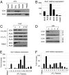
Fig. 1.
Fig. 1.
Constitutive activation of Notch in HTLV-1–infected cells. (A) Western blot analysis for expression of ICN1 in freshly isolated, uncultured samples from patients diagnosed with acute ATL. (B) Corresponding ATL patients were analyzed for hes 1 gene expression by real-time RT-PCR. (C) Half-life of ICN1 was measured by Western blot in ATL-derived cells and PBMC after cycloheximide treatment (100 μg/mL for 0, 3, and 6 h). Actin is shown as loading control. (D) Intracellular Notch is processed by receptor cleavage. Decreased ICN1 in ATL-TL–derived cells and HTLV-I–transformed C8166 cells after treatment with 1 μM GSI (Calbiochem) for 0, 2, 4, and 8 d. (E and F) Relative expression of HES1 and CDC4 mRNA, determined by real-time quantitative RT-PCR analysis, in samples from patients with acute ATL.
Fig. 2.
Fig. 2.
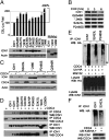
Mutations in the PEST domain of ICN1 led to its stabilization and reduced degradation by CDC4/FBW7. (A) Transcriptional activity of ICN1 and ATL ICN1 mutants using the 12XCSL luciferase reporter construct. Luciferase values were normalized and expressed compared with wild-type ICN1 set at 100%. All experiments were performed in duplicate and were repeated independently at least twice; representative data are shown as the average with SDs. Western blot showing expression of ICN1 mutants with a C-terminal Myc tag. Asterisk indicates the presence of an early termination codon at that amino acid position. *2439, *2403, and *2466 were detected with using a N-terminal Myc tag. (B) Increased half-life of several ICN1 mutants shown by Western blot analyses after cycloheximide treatment (100 μg/mL for 0, 3, and 6 h). (C) The same ATL ICN1 mutants were analyzed for CDC4-mediated degradation and compared with wild-type ICN1 after transient expression in U2OS cells in the presence of CDC4-Flag and treatment with cycloheximide (100 μg/mL for 5 h). Expression of CDC4 using Flag antibody and actin-loading controls is shown. (D). Interactions between ATL ICN1 mutants and wild-type FLAG-tagged CDC4. 293T cells were transfected with ICN1 or ICN1 mutants in the presence or absence of CDC4. IP Flag (CDC4) and WB Myc (ICN1) show interactions between ICN1, ICN1 mutants, and CDC4. ICN1 PEST domain early stop *2403 was used as negative control. WB Flag revealed equivalent amounts of CDC4 IP. Reverse IP Myc (ICN1) and WB Flag (CDC4) is shown underneath IP Myc; WB Myc is shown for control. (E) CDC4-mediated ubiquitination assay for ATL ICN1 mutants G2427S and S2423L in U2OS cells cotransfected with HA-Ub vector. Experiments were performed in the presence and absence of MG132 (10 μM). Controls for ICN1 and HA expression from lysates are shown. (F) ICN1 S2423L and wild-type ICN1 ubiquitination assay in U2OS cells transfected with HA-Ub (K48R). IP, immunoprecipitation; WB, Western blot.
Fig. 3.
Fig. 3.
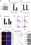
Inhibition of Notch signaling reduces proliferation and survival of ATL cells in vitro. (A) Expression of ICN1 in ATL-derived cells compared with normal PBMC. (B) Relative expression of hes1 correlates with ICN1 expression in ATL cells. Relative expression of Hes1 in T-ALL Jurkat and ATL-derived transformed cell lines [TL-Om1 and ED40515(−)] quantified by real-time quantitative RT-PCR. (C) Inhibition of Hes1 expression in ATL cells transduced with a lentiviral vector expressing DN-MAML1. (D) Inhibition of Notch signaling by DN-MAML1 results in 50–70% reduction in ATL tumor cell survival. (E) GSI-induced cell-cycle arrest in all phases of the cell cycle in ATL-derived transformed cells. For ATL-derived TL-Om1–transformed cells without GSI (TL −GSI), G1: 68.1%; S: 8.5%; G2/M: 17.9%; and for ATL-derived TL-Om1–transformed cells with GSI (TL +GSI), G1: 67.7%; S: 7.7%; G2/M: 17.2%). For ATL-derived ED40515(−)-transformed cells without GSI (ED −GSI). G1:73%; S: 11.8%; G2/M: 10.2%; and for ATL-derived ED40515(−)-transformed cells with GSI (ED +GSI), G1: 61.2%; S: 11.5%; G2/M: 12.3%. (F and G) Proliferation of ATL cells in vitro was inhibited by GSI treatment and was shown by BrdU incorporation. Quantification results represent the percentage of cells that incorporated BrdU. (H) Western blot analysis of ATL cells before and after treatment with GSI. Increased expression of CDKI p21WAF and p27KIP and cyclin B with concomitant reduction in cyclin E levels was detected after GSI-mediated inhibition of cell proliferation. Actin was used as loading control.
Fig. 4.
Fig. 4.
GSI-mediated inhibition of Notch signaling reduces ATL tumor formation in vivo. (A) Relative expression of PTEN by quantitative RT-PCR in samples from patients with acute ATL. (B) Immunodetection of cytoplasmic expression of PTEN protein in freshly isolated, uncultured ATL samples from two patients. (C) Western blot analyses showing similar expression of PTEN protein in normal PBMC and four ATL-derived transformed cells. (D) Inhibition of Notch signaling in vivo by GSI in NOG mice engrafted with ATL tumor cells. Four animals were assigned randomly to each group (not injected with ATL cells and receiving DAPT; injected with ATL cells and receiving vehicle only; or injected with ATL cells and receiving DAPT treatment). DAPT (GSI) or vehicle only was administered by oral gavage every 3 d for 28 d. One animal in group 2 injected with ATL cells and receiving vehicle only died during the 28-d period. External tumors are shown by arrows. Splenomegaly was significantly reduced in animals that received DAPT as compared with the control group. Tumors from animals were removed and measured. An ~50% reduction in tumor size was observed in animals that were treated with DAPT (mean, 78 mm3 versus 128 mm3; P < 0.05).

References

    1. D'Souza B, Miyamoto A, Weinmaster G. The many facets of Notch ligands. Oncogene. 2008;27:5148–5167. - PMC - PubMed
    1. Artavanis-Tsakonas S, Rand MD, Lake RJ. Notch signaling: Cell fate control and signal integration in development. Science. 1999;284:770–776. - PubMed
    1. Jarriault S, et al. Signalling downstream of activated mammalian Notch. Nature. 1995;377:355–358. - PubMed (V体育官网入口)
    1. Aifantis I, Raetz E, Buonamici S. Molecular pathogenesis of T-cell leukaemia and lymphoma. Nat Rev Immunol. 2008;8:380–390. - PubMed (VSports最新版本)
    1. Grabher C, von Boehmer H, Look AT. Notch 1 activation in the molecular pathogenesis of T-cell acute lymphoblastic leukaemia. Nat Rev Cancer. 2006;6:347–359. - "VSports在线直播" PubMed

Publication types

MeSH terms (VSports手机版)

Substances