Skip to main page content
U.S. flag

An official website of the United States government

Dot gov

The VSports app下载. gov means it’s official. Federal government websites often end in . gov or . mil. Before sharing sensitive information, make sure you’re on a federal government site. .

Https

The site is secure V体育官网. The https:// ensures that you are connecting to the official website and that any information you provide is encrypted and transmitted securely. .

. 2010 Sep 3;329(5996):1205-10.
doi: 10.1126/science.1192698.

The junctional adhesion molecule JAML is a costimulatory receptor for epithelial gammadelta T cell activation

Affiliations

V体育安卓版 - The junctional adhesion molecule JAML is a costimulatory receptor for epithelial gammadelta T cell activation

Deborah A Witherden et al. Science. .

VSports注册入口 - Abstract

Gammadelta T cells present in epithelial tissues provide a crucial first line of defense against environmental insults, including infection, trauma, and malignancy, yet the molecular events surrounding their activation remain poorly defined. Here we identify an epithelial gammadelta T cell-specific costimulatory molecule, junctional adhesion molecule-like protein (JAML). Binding of JAML to its ligand Coxsackie and adenovirus receptor (CAR) provides costimulation leading to cellular proliferation and cytokine and growth factor production VSports手机版. Inhibition of JAML costimulation leads to diminished gammadelta T cell activation and delayed wound closure akin to that seen in the absence of gammadelta T cells. Our results identify JAML as a crucial component of epithelial gammadelta T cell biology and have broader implications for CAR and JAML in tissue homeostasis and repair. .


Figures

Fig. 1
Fig. 1
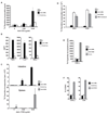
Murine JAML, a 55kDa protein expressed on γδ T cells, is recognized by mAb HL4E10. (A) 7–17 cells were analyzed by flow cytometry for HL4E10 or control mAb staining. (B) γδ TCR and HL4E10 fluorescent staining (magnification ×600) of 7–17 cells grown on coverslips. The nucleus is stained with DAPI. Scale bar represents 50µm. (C) Western blot analysis of 7–17 lysates immunoprecipitated with mAb HL4E10 (lane 1) or a control hamster mAb, 1F4, (lane 2). Protein identification by LC-ESI-MS gave a high scoring match with mouse JAML (Mascot score 151). (D) Flow cytometric analysis of JAML expression by resting and ConA activated epithelial and peripheral γδ T cells. Cells were gated on γδ TCR+. (E) Quantitative RT-PCR analysis of JAML expression in γδ T cells from epidermis, intestine and spleen. Relative amounts of JAML mRNA were normalized to β-actin and are shown as arbitrary units. (F) Flow cytometric analysis of JAML expression by splenic B cells, CD4+ T cells and CD8+ T cells. Both resting and activated B cells (LPS-activated) and αβ T cells (PMA and ionomycin) were analyzed. (G) Quantitative RT-PCR analysis of JAML expression in CD4+CD8 and CD4CD8+ splenocytes. Relative amounts of JAML mRNA were normalized to β-actin and are shown as arbitrary units. ND=not determined. (H) JAML mRNA isolated from representative epithelial and lymphocyte cell lines: PAM keratinocytes (lane 1), 3T3 fibroblasts (lane 2), DP thymocytes (DPK; lane 3), CD4+ T cells (D10; lane 4), CD8+ T cells (CTLL; lane 5), B cells (A20; lane 6), γδ T cells (7–17; lane 7) and RT control (lane 8). Data in (A) through (H) are representative of at least three experiments.
Fig. 2
Fig. 2
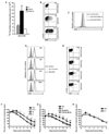
JAML functions as a costimulatory molecule on epithelial γδ T cells, but not lymphoid γδ T cells or αβ T cells. (A) Proliferation of short-term DETC cell lines to immobilized anti-CD3 either alone or in combination with anti-JAML mAb, HL4E10, or a control mAb, 1F4. (B) JAML costimulation induces cytokine production. ELISA analysis for TNFα, IL-2 and IFNγ from culture supernatants of DETC stimulated with anti-CD3 alone, anti-CD3 and anti-JAML, or anti-CD3 and control mAb, 1F4. (C) Proliferation of intestinal or splenic γδ T cells to immobilized anti-γδ TCR mAb (GL3) either alone or in combination with anti-JAML, control mAb 1F4, or anti-CD28. (D) Proliferation of splenic CD4+ and CD8+ T cells in response to immobilized anti-CD3 (0.4µg/ml) alone or in combination with anti-JAML or anti-CD28. PMA and ionomycin was used as a positive control. (E) Proliferation of sorted CD8αα+ αβ IEL to immobilized anti-CD3 (0.4µg/ml) alone or in combination with anti-JAML or anti-CD28. Con A was used as a positive control. (F) IL-2 production by CD8αβ+ and CD4+ αβ IEL in response to anti-CD3 (0.4µg/ml) in combination with anti-JAML, control mAb 1F4, or anti-CD28. IL-2 was measured by MTT assay. Data in (A) through (F) are representative of at least three experiments.
Fig. 3
Fig. 3
CAR is a functional ligand for JAML in the epidermis. (A) PAM 2–12 and PDV keratinocyte and 3T3 fibroblast cell lines were analyzed by flow cytometry for binding of soluble JAML (sJAML), anti-CD29 (positive control) or streptavidin PE (SA-PE; negative control). (B) Western blot analysis of PDV lysates immunoprecipitated with sJAML (lane 1) or empty beads (lane 2). (C–F) PDV keratinocytes were analyzed by flow cytometry for binding of sJAML and soluble adenovirus fiber protein (F5). Keratinocytes were stained with sJAML or F5 alone (C), sJAML followed by F5 (D), F5 followed by sJAML (E), or stained with sJAML, fixed and then stained with F5 (F). (G) Proliferation of the 7–17 γδ T cell line to immobilized anti-CD3 either alone or in combination with CAR-Fc, a negative control Fc fusion protein, CD62L-Fc, HL4E10, or control mAb 1F4. (H) Western blot analysis of phosphorylation of Akt Ser473 and Thr308 and total Akt in 7–17 DETC following JAML ligation with CAR-Fc, in the presence or absence (DMSO vehicle control) of the PI3K inhibitor LY924002. β-actin was used as a loading control. (I,J) Western blot analysis of ERK 1,2 phosphorylation and p38 and JNK kinase activity in 7–17 lysates following 5 min stimulation with anti-CD3, anti-JAML or both. Total ERK is shown as a control. Quantification of data in (I) is shown in (J). Data in (A) through (J) are representative of at least three experiments.
Fig. 4
Fig. 4
Costimulation through JAML-CAR interactions mediates epithelial γδ T cell activation and effector function. (A) Proliferation of freshly isolated epidermal γδ T cells to immobilized anti-CD3 (1µg/ml) either alone (0) or in combination with CAR-Fc or a negative control Fc fusion protein, CD62L-Fc. (B) Intracellular flow cytometric analysis of TNFα production by unstimulated, anti-CD3 stimulated (0.5µg/ml) or anti-CD3 (0.05µg/ml) and CAR-Fc (10µg/ml) stimulated 7–17 DETC. (C) Flow cytometric analysis of KGF expression by unstimulated primary DETC or after stimulation with immobilized anti-CD3 (1µg/ml) and CAR-Fc or anti-CD3 and control Fc fusion protein, CD62L-Fc. (D) Flow cytometric analysis of CAR expression by keratinocytes isolated from epidermis of C57Bl/6 mice. Skin was either non-wounded or wounded and keratinocytes were isolated at the indicated times. (E) Flow cytometric analysis of TNFα production by DETC isolated from epidermis of C57Bl/6 mice. Skin was either non-wounded, wounded and treated with soluble F5 protein (F5-treated), wounded and treated with control soluble F11 protein (Ctrl-treated) or wounded and not treated. (F) Wound closure in vivo in the presence of soluble F5 or a control protein, F11. Untreated C57Bl/6 mice (0) and TCRδ−/− mice (Δ) were used as controls. (G) Wound closure in the presence of F5, HL4E10 or F5 and HL4E10. Untreated C57Bl/6 mice (0) were used as a control for rate of wound closure. (H) Wound closure in F5-treated (F5) and untreated (0) TCRδ−/− mice. Data represent mean ± SEM of 4–8 wounds per condition. Student’s t test p values (**p<0.005 and *p<0.05). Data in (A) through (H) are representative of more than three experiments.

V体育平台登录 - Comment in

  • Immunology. CAR'ing for the skin.
    Shaw AS, Huang Y. Shaw AS, et al. Science. 2010 Sep 3;329(5996):1154-5. doi: 10.1126/science.1195337. Science. 2010. PMID: 20813941 No abstract available.

V体育安卓版 - References

    1. Havran WL, Chien YH, Allison JP. Recognition of self antigens by skin-derived T cells with invariant γδ antigen receptors. Science. 1991;252:1430. - PubMed
    1. Jameson J, et al. A role for skin γδ T cells in wound repair. Science. 2002;296:747. - PubMed
    1. Strid J, Tigelaar RE, Hayday AC. Skin immune surveillance by T cells-a new order? Semin. Immunol. 2009;21:110. - PubMed
    1. Girardi M, et al. Regulation of cutaneous malignancy by γδ T cells. Science. 2001;294:605. - PubMed
    1. Sharp LL, Jameson JM, Cauvi G, Havran WL. Dendritic epidermal T cells regulate skin homeostasis through local production of insulin-like growth factor 1. Nat. Immunol. 2005;6:73. - PubMed

Publication types (VSports注册入口)

MeSH terms

Substances