VSports在线直播 - Skip to main page content
U.S. flag

An official website of the United States government

Dot gov

The . gov means it’s official. Federal government websites often end in . gov or VSports app下载. mil. Before sharing sensitive information, make sure you’re on a federal government site. .

Https

The site is secure V体育官网. The https:// ensures that you are connecting to the official website and that any information you provide is encrypted and transmitted securely. .

. 2010 Aug 18;5(8):e12238.
doi: 10.1371/journal.pone.0012238.

Bacteria penetrate the inner mucus layer before inflammation in the dextran sulfate colitis model

Affiliations

"V体育2025版" Bacteria penetrate the inner mucus layer before inflammation in the dextran sulfate colitis model

VSports最新版本 - Malin E V Johansson et al. PLoS One. .

V体育官网入口 - Abstract

Background: Protection of the large intestine with its enormous amount of commensal bacteria is a challenge that became easier to understand when we recently could describe that colon has an inner attached mucus layer devoid of bacteria (Johansson et al. (2008) Proc. Natl. Acad. Sci. USA 105, 15064-15069). The bacteria are thus kept at a distance from the epithelial cells and lack of this layer, as in Muc2-null mice, allow bacteria to contact the epithelium. This causes colitis and later on colon cancer, similar to the human disease Ulcerative Colitis, a disease that still lacks a pathogenetic explanation. Dextran Sulfate (DSS) in the drinking water is the most widely used animal model for experimental colitis. In this model, the inflammation is observed after 3-5 days, but early events explaining why DSS causes this has not been described VSports手机版. .

Principal findings: When mucus formed on top of colon explant cultures were exposed to 3% DSS, the thickness of the inner mucus layer decreased and became permeable to 2 microm fluorescent beads after 15 min. Both DSS and Dextran readily penetrated the mucus, but Dextran had no effect on thickness or permeability. When DSS was given in the drinking water to mice and the colon was stained for bacteria and the Muc2 mucin, bacteria were shown to penetrate the inner mucus layer and reach the epithelial cells already within 12 hours, long before any infiltration of inflammatory cells. V体育安卓版.

Conclusion: DSS thus causes quick alterations in the inner colon mucus layer that makes it permeable to bacteria. The bacteria that reach the epithelial cells probably trigger an inflammatory reaction. These observations suggest that altered properties or lack of the inner colon mucus layer may be an initial event in the development of colitis. V体育ios版.


"VSports在线直播" Conflict of interest statement

Competing Interests: The authors have declared that no competing interests exist.

Figures (VSports在线直播)

Figure 1
Figure 1. Direct effects of Dextran Sulfate (DSS) on mucus formed by explant cultures of human and mouse colon.
(A) Effects of 3% DSS and 3% Dextran on the mucus thickness in mouse distal colon explants (n = 7 in each group, p<0.001) and human sigmoid colon biopsies (n = 6 in each group, p<0.01). Data is presented as mean ± SEM. The control values are normalized to 100 and the student's t-test was used to analyze the effect of the respective treatments. (B) The tissue showed a nice crypt architecture in the XY focal plane when stained with CellTracer BODIPY TR methyl ester as seen in red. (C) Z-section of an X-Y confocal image stack of secreted mucus on a mouse colon explants exposed to 3%DSS or Dextran for 15 min. The tissue was stained with a red fluorescent dye and 2 µm green fluorescent beads were left to sediment onto the mucus. During control conditions the beads remained on top of the mucus. Exposure to DSS resulted in reduced mucus thickness and beads were able to reach the epithelial surface within 15 min. Dextran had no effect on either mucus thickness or permeability. Mucus top surface (1), inner firm mucus (2), and epithelium (3) are marked to the left and right.
Figure 2
Figure 2. Histology of mouse colon stained with Haematoxylin/Eosin for different times of DSS exposure.
No increase in the amount of infiltrating leukocytes or altered epithelial architecture as sign for inflammation was observed after 12 and 24 h of DSS treatment. Inflammation with infiltrating leukocytes and loss of normal epithelial architecture was obvious after 120 h of DSS administration. Scale bars are 100 µm.
Figure 3
Figure 3. The epithelium can produce a normal mucus layer after 24 h of DSS treatment.
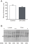
(A) The thickness of the inner firm mucus layer was measured in vivo in mice after removal of the outer loose mucus layer. The measurements were made in control animals (n = 6) and in mice treated with DSS for 24 h (n = 3). No significant difference was observed. (B) The loose and firm mucus from animals treated with DSS for 12, 18 and 24 h were reduced and separated by AgPAGE and stained with Alcian blue. The Muc2 bands migrate at their normal position for a monomer (M) and non-reducible oligomers.
Figure 4
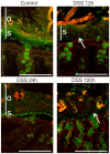
Figure 4. Localization of bacteria in the colon mucus of mice after DSS treatments for 12, 24 and 120 h or in nontreated control (No DSS).
Bacteria were stained by fluorescent in-situ hybridization using the general bacterial rRNA probe, EUB338 conjugated with Alexa 555 (red). The mucus was visualized by immunostaining of the same section with an anti-Muc2 specific antiserum (green). Penetration of bacteria through the inner firm and stratified (s) mucus layer was observed already after 12 h DSS administration. The inner stratified mucus layer is marked by s and the outer by o when any of the layers could be identified. Arrows point out bacteria within the inner mucus layer (12 h) or close to the epithelium in the absence of an inner mucus layer (120 h). Scale bars are 100 µm.
Figure 5
Figure 5. High number of bacteria penetrating the inner mucus layer co-varies with high amounts of DSS in the colon mucus.
(A) Scoring of bacteria penetration of the inner firm mucus layer on a scale from 0 (no penetration) to 5 (large number of bacteria reaching the epithelial surface) of controls and DSS treated mice for indicated times. The scoring system is exemplified and demonstrated in Supplement Fig. S1. Mean values are for three mice (one mouse at 120 h) scored at four sites by two individuals in a blinded fashion. (B) Mucus from the analyzed animals was extracted with guanidinium chloride, the soluble fraction was reduced, separated on AgPAGE, and the gel stained with Alcian blue. The Muc2 monomer is marked by M. The DSS was also stained and is as expected absent in the untreated control animals. The amount of DSS reflects the animals diurnal activity and drinking rhythm as 12 and 36 h are after a night of activity and thus show high levels of DSS compared to 18 and 24 h that are after an inactive daytime that show low levels of DSS.
Figure 6
Figure 6. DSS treatment of mice allowed bacteria to penetrate into the firm mucus layer and come in direct contact with the epithelium.
(A) Mice were given FITC-DSS (3%, green) in the drinking water for 12 h. The distal colon was fixed and the sections were immunostained with anti-Muc2 antiserum and anti-rabbit-Alexa546 (red). The arrows point at FITC-DSS that has penetrated the inner stratified mucus layer (s). Large amounts of bacteria and FITC-DSS is shown in the outer loose mucus layer (o). Epithelial cells are marked (e). Scale bar is 25 µm. (B) Bacteria were stained with FISH using the general rRNA probe EUB338 conjugated to Alexa 555 (red) and the nuclear DNA stained with Sytox Green DNA stain (green). The picture shows massive amounts of bacteria in contact with the epithelial cells. Scale bar is 10 µm.

Comment in

References

    1. Atuma C, Strugula V, Allen A, Holm L. The adherent gastrointestinal mucus gel layer: thickness and physical state in vivo. Am J Physiol Gastrointest Liver Physiol. 2001;280:G922–G929. - PubMed
    1. Johansson MEV, Phillipson M, Petersson J, Holm L, Velcich A, Hansson GC. The inner of the two Muc2 mucin dependent mucus layers in colon is devoid of bacteria. Proc Natl Acad Sci USA. 2008;105:15064–15069. - PMC (V体育官网) - PubMed
    1. Lang T, Hansson GC, Samuelsson T. Gel-forming mucins appeared early in metazoan evolution. Proc Natl Acad Sci U S A. 2007;104:16209–16214. - PMC - PubMed
    1. Johansson MEV, Thomsson KA, Hansson GC. Proteomic Analyses of the Two Mucus Layers of the Colon Barrier Reveal That Their Main Component, the Muc2 Mucin, Is Strongly Bound to the Fcgbp Protein. J Proteome Res. 2009;8:3549–3557. - PubMed
    1. Asker N, Axelsson MAB, Olofsson SO, Hansson GC. Dimerization of the human MUC2 mucin in the endoplasmic reticulum is followed by a N-glycosylation-dependent transfer of the mono- and dimers to the Golgi apparatus. J Biol Chem. 1998;273:18857–18863. - V体育平台登录 - PubMed

MeSH terms

"V体育安卓版" Substances

VSports app下载 - Grants and funding