Skip to main page content
U.S. flag

An official website of the United States government

Dot gov

The . gov means it’s official. Federal government websites often end in . gov or VSports app下载. mil. Before sharing sensitive information, make sure you’re on a federal government site. .

Https

The site is secure V体育官网. The https:// ensures that you are connecting to the official website and that any information you provide is encrypted and transmitted securely. .

. 2010 Oct 1;185(7):4030-41.
doi: 10.4049/jimmunol.1001778. Epub 2010 Aug 30.

Impaired phagocytosis of apoptotic cells by macrophages in chronic granulomatous disease is reversed by IFN-γ in a nitric oxide-dependent manner (V体育2025版)

Affiliations

Impaired phagocytosis of apoptotic cells by macrophages in chronic granulomatous disease is reversed by IFN-γ in a nitric oxide-dependent manner

Ruby Fernandez-Boyanapalli et al. J Immunol. .

Abstract

Immunodeficiency in chronic granulomatous disease (CGD) is well characterized. Less understood are exaggerated sterile inflammation and autoimmunity associated with CGD. Impaired recognition and clearance of apoptotic cells resulting in their disintegration may contribute to CGD inflammation. We hypothesized that priming of macrophages (Ms) with IFN-γ would enhance impaired engulfment of apoptotic cells in CGD. Diverse M populations from CGD (gp91(phox)(-/-)) and wild-type mice, as well as human Ms differentiated from monocytes and promyelocytic leukemia PLB-985 cells (with and without mutation of the gp91(phox)), demonstrated enhanced engulfment of apoptotic cells in response to IFN-γ priming. Priming with IFN-γ was also associated with increased uptake of Ig-opsonized targets, latex beads, and fluid phase markers, and it was accompanied by activation of the Rho GTPase Rac. Enhanced Rac activation and phagocytosis following IFN-γ priming were dependent on NO production via inducible NO synthase and activation of protein kinase G. Notably, endogenous production of TNF-α in response to IFN-γ priming was critically required for inducible NO synthase upregulation, NO production, Rac activation, and enhanced phagocytosis. Treatment of CGD mice with IFN-γ also enhanced uptake of apoptotic cells by M in vivo via the signaling pathway. Importantly, during acute sterile peritonitis, IFN-γ treatment reduced excess accumulation of apoptotic neutrophils and enhanced phagocytosis by CGD Ms. These data support the hypothesis that in addition to correcting immunodeficiency in CGD, IFN-γ priming of Ms restores clearance of apoptotic cells and may thereby contribute to resolution of exaggerated CGD inflammation VSports手机版. .


"V体育平台登录" Figures
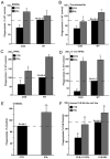
Figure 1
Figure 1
IFN-γ priming of CGD and WT Mϕ populations in vitro and in vivo enhances uptake of apoptotic cells. BMDMϕs (A), thioglycollateelicited Mϕs (B), RPMϕs (C), HMDMϕs (E), and Mϕ-like differentiated PLB-985 and mutated PLB-985 (X-CGD) cell lines (F) were primed overnight with IFN-γ (50 ng/ml, 4200 U) or PBS in vitro and uptake of apoptotic Jurkat cells was assayed (n = 5 for each condition). D, Mice were treated with IFN-γ (500 ng, 42,000 U) or PBS i.p. and 24 h later, PKH-labeled apoptotic thymocytes were instilled into peritonea and in vivo uptake by Mϕs was assessed (see Materials and Methods) (n = 4). Dashed line represents baseline uptake by normal or WT Mϕs, and PI is shown for each cell type; *p ≤ 0.05 compared with control Mϕs of each cell type, respectively, and αp ≤ 0.05 compared with uptake by normal or WT Mϕs of each cell type, respectively, at baseline. PI, phagocytic index; RPMϕ, resident peritoneal Mϕ.
Figure 2
Figure 2
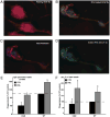
IFN-γ priming enhances ruffling, fluid phase uptake, and phagocytosis of opsonized viable cells and latex beads by Mϕs. Resting (A) and IFN-γ primed (50 ng/ml, overnight) (BD) RAW 264.7 cells were analyzed for actin polymerization (red, C) and uptake of LY dye (green, D) at ×100 magnification. Yellow indicates colocalization of markers (B). BMDMϕs were primed with IFN-γ or not as in Fig. 1, and the uptake of Ig-opsonized Jurkat cells (E) (n = 3) or latex beads (5 μm) (F) (n = 3) was assayed. *p ≤ 0.05 compared with respective control cells.
Figure 3
Figure 3
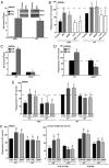
Enhanced phagocytosis of apoptotic cells by IFN-γ–primed CGD and WT Mϕs is dependent on NO production. IFN-γ priming of BMDMϕs was performed as in Fig. 1. A, iNOS was detected by immunobloting (inset) and NO was measured in supernatants following overnight culture (n = 3). B, Cells were IFN-γ primed (or not) or SNAP treated (50 μM for 24 h) and, where indicated, cells were treated with L-NIL (0.5 mM) for 30 min before phagocytosis assayed (n = 3). BMDMϕs from WT and iNOS−/− mice were primed with IFN-γ and NO production (C) or uptake of apoptotic cells (D) was determined (n = 5). E, Cells were treated with the PKG inhibitor, KT5823 (10 μM for 30 min) before IFN-γ priming or treated with SNAP as in B, and uptake of apoptotic cells was determined for BMDMϕs (n = 5) (E), HMDMϕs (n = 10), and the PLB-985 Mϕ cell lines (n = 4) (F). *p ≤ 0.05 compared with the untreated control Mϕs for each cell type, respectively; αp ≤ 0.04 compared with IFN-γ–primed Mϕs of each cell type, respectively; #p ≤ 0.04 compared with SNAP-treated Mϕs of each cell type, respectively; δp ≤ 0.03 compared with IFN-γ–primed WT Mϕs.
Figure 4
Figure 4
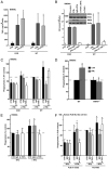
TNF-α production and action in response to IFN-γ priming is required for iNOS induction and NO production. A, TNF-α was measured in the supernatants of BMDMϕs following IFN-γ priming with and without treatment with the PKG inhibitor KT5823 as in Fig. 3 (n = 5). B, BMDMϕs were primed with IFN-γ in the presence of A-TNF-α or isotype control and NO was measured in supernatants and iNOS was detected by immunoblotting (inset, one representative experiment). Following treatment as in B, phagocytosis of apoptotic cells was determined in Mϕs: BMDMϕs from WT and CGD mice (n = 5) (C), WT and TNFR1−/− mice (D) (n = 5), HMDMϕs (n = 10) (E), and PLB-X-CGD and PLB-985 cell lines (n = 4) (F). *p ≤ 0.04 compared with the untreated control Mϕs for each cell type, respectively; αp ≤ 0.04 compared with IFN-γ–primed Mϕs of each cell type, respectively; #p ≤ 0.04 compared with SNAP-treated Mϕs of each cell type, respectively; δp ≤ 0.02 compared with IFN-γ–primed WT Mϕs. A-TNF-α, neutralizing Ab to TNF-α.
Figure 5
Figure 5
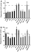
IFN-γ priming enhances phagocytosis of Ig-opsonized Jurkat cells by the TNF-α/iNOS/NO-PKG–dependent mechanism. BMDMϕs from CGD (A) and WT (B) mice were treated with the agents/inhibitors as previously described, and phagocytosis of CD45-opsonized Jurkat cells was determined (n = 4). *p ≤ 0.05 compared with the control Mϕs for each cell type, respectively; αp ≤ 0.05 compared with IFN-γ–primed Mϕs of each cell type, respectively; #p ≤ 0.02 compared with SNAP-treated Mϕ of each cell type, respectively.
Figure 6
Figure 6
IFN-γ priming leads to Rac activation by the TNF-α/NO/PKG–dependent pathway. BMDMϕs from CGD (A) and WT (B) mice were tested for Rac activation using the agents/inhibitors as in Fig. 3, and representative immunoblots are shown. C, Densitometry was used to determine the ratio of active Rac to total Rac (n = 3). *p ≤ 0.05 compared with the control Mϕs for each cell type, respectively; αp ≤ 0.05 compared with IFN-γ–primed Mϕs of each cell type, respectively; #p ≤ 0.05 compared with SNAP-treated Mϕs of each cell type, respectively.
Figure 7
Figure 7
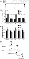
In vivo treatment with IFN-γ enhances ex vivo phagocytosis of apoptotic cells by resident peritoneal Mϕs via the TNF-α/NO/PKG pathway. A, Experimental design is shown. CGD (B) and WT (C) mice were injected i.p. with IFN-γ or PBS as in Fig. 1D; 24 h later peritoneal Mϕs were lavaged, plated, rested for 24 h in the presence or absence of the specified inhibitors/agents, and then phagocytosis of apoptotic Jurkat cells was assessed (n = 5). Dashed line represents uptake in WT Mϕs following PBS treatment. *p ≤ 0.05 compared with control mice of each genotype injected with PBS, respectively; #p ≤ 0.05 compared with mice of each genotype injected with IFN-γ, respectively. D, Mechanism of IFN-γ enhancement of uptake operant both in vitro and ex vivo.
Figure 8
Figure 8
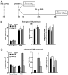
IFN-γ treatment of CGD mice enhances phagocytosis of apoptotic cells by Mϕs during resolution of inflammation. A, Experimental design is shown. CGD and WT mice were injected i.p. with IFN-γ (500 ng, 42,000 U) or PBS and 24 h later all mice were injected with 1 mg of zymosan i.p. B, Mice in subgroup I (6 h after zymosan treatment) were euthanized and peritoneal cells lavaged and analyzed for differentials, apoptotic neutrophils, and phagocytic indices. C, Mice in subgroup II were treated with a second dose of PBS or IFN-γ at 24 h after zymosan treatment, and peritoneal cells were analyzed at 48 h after zymosan treatment. *p ≤ 0.03 compared with mice treated with PBS for each genotype, respectively; αp ≤ 0.04 compared with WT mice treated with PBS.

References

    1. Gallin JI, Buescher ES. Abnormal regulation of inflammatory skin responses in male patients with chronic granulomatous disease. Inflammation. 1983;7:227–232. - PubMed
    1. Romani L, Fallarino F, De Luca A, Montagnoli C, D'Angelo C, Zelante T, Vacca C, Bistoni F, Fioretti MC, Grohmann U, et al. Defective tryptophan catabolism underlies inflammation in mouse chronic granulomatous disease. Nature. 2008;451:211–215. - PubMed
    1. Segal BH, Leto TL, Gallin JI, Malech HL, Holland SM. Genetic, biochemical, and clinical features of chronic granulomatous disease. Medicine (Baltimore) 2000;79:170–200. - PubMed
    1. Winkelstein JA, Marino MC, Johnston RB, Jr, Boyle J, Curnutte J, Gallin JI, Malech HL, Holland SM, Ochs H, Quie P, et al. Chronic granulomatous disease: report on a national registry of 368 patients. Medicine (Baltimore) 2000;79:155–169. - PubMed
    1. Holland SM. Chronic granulomatous disease. Clin Rev Allergy Immunol. 2010;38:3–10. - PubMed (V体育2025版)

"V体育2025版" Publication types

MeSH terms