<acronym id="zvkyo"></acronym>
"VSports app下载" Skip to main page content
U.S. flag

An official website of the United States government

Dot gov

The . gov means it’s official. Federal government websites often end in . gov or VSports app下载. mil. Before sharing sensitive information, make sure you’re on a federal government site. .

Https

The site is secure. The https:// ensures that you are connecting to the official website and that any information you provide is encrypted and transmitted securely V体育官网. .

. 2010 Sep 1;185(5):2670-4.
doi: 10.4049/jimmunol.1001610. Epub 2010 Aug 2.

V体育安卓版 - Cutting edge: mutation of Francisella tularensis mviN leads to increased macrophage absent in melanoma 2 inflammasome activation and a loss of virulence

Affiliations

Cutting edge: mutation of Francisella tularensis mviN leads to increased macrophage absent in melanoma 2 inflammasome activation and a loss of virulence (VSports在线直播)

Tyler K Ulland et al. J Immunol. .

Abstract

The mechanisms by which the intracellular pathogen Francisella tularensis evades innate immunity are not well defined. We have identified a gene with homology to Escherichia coli mviN, a putative lipid II flippase, which F. tularensis uses to evade activation of innate immune pathways. Infection of mice with a F. tularensis mviN mutant resulted in improved survival and decreased bacterial burdens compared to infection with wild-type F. tularensis. The mviN mutant also induced increased absent in melanoma 2 inflammasome-dependent IL-1beta secretion and cytotoxicity in macrophages. The compromised in vivo virulence of the mviN mutant depended upon inflammasome activation, as caspase 1- and apoptosis-associated speck-like protein containing a caspase recruitment domain-deficient mice did not exhibit preferential survival following infection. This study demonstrates that mviN limits F. tularensis-induced absent in melanoma 2 inflammasome activation, which is critical for its virulence in vivo VSports手机版. .


Figures

Figure 1
Figure 1
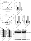
F. tularensis LVS mviN mutant is attenuated in vivo. (A) WT (n=10 per group) mice were injected i.p. with the indicated amount of F. tularensis LVS (LVS) or the mviN mutant (mviN::Tn5) and survival monitored. Data are representative of 5 independent experiments at the 3 × 10⁴ infective dose, each involving a minimum of 5 mice per group. (B) 3 d p.i. with 3 × 10⁴ CFU of LVS or the mviN mutant organs were harvested, homogenized and dilutions plated for enumeration of CFU (n=3–5 mice per group). (C) Number of lesions per 200 × field of H&E-stained liver sections from WT mice 3 d p.i. with either LVS or the mviN mutant were scored; n=5 mice per group (3 random fields were examined per mouse); *, p = 0.016. (D) Representative H&E-stained sections of liver from WT mice 3 d p.i. with either F. tularensis LVS or the mviN mutant. Arrows indicate necropurulent foci.
Figure 2
Figure 2
Mutation of F. tularensis LVS mviN results in enhanced Mφ cytotoxicity and caspase-1 activation. (A–E) LPS-primed WT Mφ were infected with F. tularensis LVS (LVS), the mviN mutant (mviN::Tn5) or the complemented mviN mutant (mviN::Tn5 + mviN) at an MOI of 50:1. Supernatants were collected 9 h after infection (B, D, and E) or as indicated (A and C). Cytotoxicity was measured by LDH release and expressed as a percentage of LDH release by Triton X-100 detergent (A and B). IL-1β secreted into supernatants was measured by ELISA (C-E). (F, G) Unprimed WT Mφ were infected with LVS or the mviN mutant for 9 h; IL-6 and TNFα released into the supernatant was measured by ELISA. (H) Lysates from LPS-primed WT and caspase-1-deficient Mφ (lane with caspase-1-/- Mφ lysate is marked *) infected with LVS or the mviN mutant (50:1 MOI) for the indicated times were immunoblotted with antibodies against the p10 subunit of caspase-1 or GAPDH. Results are representative of two (B, F, G) and three (A, C-E, H) separate experiments.
Figure 3
Figure 3
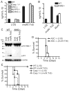
MviN limits Francisella-induced AIM2 inflammasome activation. (A, B) LPS-primed Mφ from WT, caspase-1-, ASC-, NLRP3-, NLRC4, and AIM2-deficient mice were infected with either F. tularensis LVS (LVS) or the mviN mutant (mviN::Tn5) at a 50:1 MOI, or challenged with Alum (500 μg/ml). Supernatants were collected at 9 h and IL-1β release measured by ELISA. Results are representative of two (A) and five (B) separate experiments. (C) Lysates from LPS-primed WT and AIM2-deficient Mφ infected with LVS or the mviN mutant for 9 h were immunoblotted with antibodies against the p10 subunit of caspase-1 or GAPDH. Results are representative of two separate experiments. (D, E) WT, caspase-1- or ASC-deficient mice (n=5 per group) were infected i.p. with 1 × 10⁵ CFUs of LVS or the mviN mutant and survival monitored.

References (VSports在线直播)

    1. Clemens DL, Lee BY, Horwitz MA. Virulent and avirulent strains of Francisella tularensis prevent acidification and maturation of their phagosomes and escape into the cytoplasm in human macrophages. Infect Immun. 2004;72:3204–3217. - "VSports app下载" PMC - PubMed
    1. Checroun C, Wehrly TD, Fischer ER, Hayes SF, Celli J. Autophagy-mediated reentry of Francisella tularensis into the endocytic compartment after cytoplasmic replication. Proc Natl Acad Sci U S A. 2006;103:14578–14583. - PMC - PubMed
    1. Telepnev M, Golovliov I, Grundstrom T, Tarnvik A, Sjostedt A. Francisella tularensis inhibits Toll-like receptor-mediated activation of intracellular signalling and secretion of TNFα and IL-1 from murine macrophages. Cell Microbiol. 2003;5:41–51. - PubMed
    1. McCaffrey RL, Allen LA. Francisella tularensis LVS evades killing by human neutrophils via inhibition of the respiratory burst and phagosome escape. J Leukoc Biol. 2006;80:1224–1230. - PMC - PubMed
    1. Mariathasan S, Weiss DS, Dixit VM, Monack DM. Innate immunity against Francisella tularensis is dependent on the ASC/caspase-1 axis. J Exp Med. 2005;202:1043–1049. - PMC - PubMed

Publication types

MeSH terms