Skip to main page content
U.S. flag

An official website of the United States government

Dot gov

The . gov means it’s official. Federal government websites often end in . gov or . mil. Before sharing sensitive information, make sure you’re on a federal government site VSports app下载. .

Https

The site is secure. The https:// ensures that you are connecting to the official website and that any information you provide is encrypted and transmitted securely. V体育官网.

. 2010 Aug 6;142(3):409-19.
doi: 10.1016/j.cell.2010.06.040.

A large intergenic noncoding RNA induced by p53 mediates global gene repression in the p53 response

Affiliations

A large intergenic noncoding RNA induced by p53 mediates global gene repression in the p53 response

Maite Huarte et al. Cell. .

"VSports注册入口" Abstract

Recently, more than 1000 large intergenic noncoding RNAs (lincRNAs) have been reported. These RNAs are evolutionarily conserved in mammalian genomes and thus presumably function in diverse biological processes VSports手机版. Here, we report the identification of lincRNAs that are regulated by p53. One of these lincRNAs (lincRNA-p21) serves as a repressor in p53-dependent transcriptional responses. Inhibition of lincRNA-p21 affects the expression of hundreds of gene targets enriched for genes normally repressed by p53. The observed transcriptional repression by lincRNA-p21 is mediated through the physical association with hnRNP-K. This interaction is required for proper genomic localization of hnRNP-K at repressed genes and regulation of p53 mediates apoptosis. We propose a model whereby transcription factors activate lincRNAs that serve as key repressors by physically associating with repressive complexes and modulate their localization to sets of previously active genes. .


Figures (V体育平台登录)

Figure 1
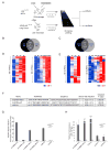
Figure 1. Several lincRNAs are p53 transcriptional targets
(A). Experiment layout to monitor p53-dependent transcription. p53-restored (+Cre) or not restored (-Cre) p53LSL/LSL MEFs were treated with 500nM dox for 0, 3, 6 and 9 hours (top left). KRAS (p53LSL/LSL) tumor cells were treated with hydroxytamoxifen for p53 restoration for 0, 8, 16, 24, 40 or 48 hours (bottom left). RNA was subjected to microarray analysis of mRNAs and lincRNAs. (B) and (C). Venn diagrams showing the number of shared and distinct mRNAs (B) or lincRNAs (C) induced in a p53-dependent manner in the MEF or KRAS systems. (D) and (E). mRNAs (D) and lincRNAs (E) activated by p53 induction (FDR < 0.05) in MEF or KRAS system. Colors represent transcripts above (red) or below (blue) the global median scaled to 8 fold activation or repression, respectively. (F) Promoter region, conserved p53 binding motif, promoter orientation and p53-dependent fold induction in reporter assays of lincRNA promoters induced in a p53-dependent manner (values are average of at least three biological replicates (+/-STD); P values are determined by t-test) (G) p53-dependent induction of lincRNA promoters requires the consensus p53 binding elements. Relative firefly luciferase expression driven by promoters with p53 consensus motif (lincRNA-p21, lincRNA-Mkln1) or with deleted motif (ΔlincRNA-p21 and ΔlincRNA-Mkln1) in p53-restored p53LSL/LSL (p53+/+) or p53LSL/LSL (p53-/-) cells. Values are relative to p53-/- and normalized by renilla levels (average of 3 replicates +/-STD). (H) p53 specifically binds to p53 motifs in lincRNA promoters. p53 ChIP enrichment in p53+/+ and p53-/- MEFs on regions with p53 motifs (lincRNA-p21, lincRNA-Mkln1, Cdkn1a) or two irrelevant regions (controls). Enrichment values are relative to IgG and average of 3 replicates (+/-STD). See also Figure S1 and Table S1.
Figure 2
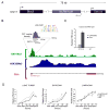
Figure 2. lincRNA-p21: a new p53 target gene induced in different tumor models
(A) Schematic representation of the chromosomal location of the lincRNA-p21 gene locus. Arrowheads indicate the orientation of transcription. (B) Promoter and transcript structure of lincRNA-p21 gene locus. Chromatin structure at the lincRNA -p21 locus is shown as mES ChIP-Seq data (Mikkelsen et al., 2007); for each histone modification (H3K4me3, green; H3K36me3, blue), ChIP-seq results are plotted as number of DNA fragments obtained at each position relative to the genomic average. Red stars indicate the position of the p53-binding motif. The promoter region where p53 ChIP-PET fragments (black segments) map is enlarged (Wei et al., 2006). PET overlap density (grey) and p53 motif sequence are shown. The structure of the full-length lincRNA-p21 is represented with red boxes as exons and arrowed lines as the intronic sequence. (C) Human lincRNA-p21 is induced by DNA damage. Relative RNA levels of human lincRNA-p21 determined by qRT-PCR (RT-PCR) or qPCR (-RT) from untreated human fibroblasts or 500nM DOX-treated for 14 hours. PCR primers map on the human region orthologus to the first exon of the mouse gene. (D) lincRNA-p21 is induced by p53 in different tumor cell lines. lincRNA-p21, p53 and Cdkn1a relative RNA levels at different times after p53 restoration. Values are the median of 4 technical replicates. Values in (C) and (D) are the median of 4 technical replicates (+/-STD). See also Figure S2.
Fig 3
Fig 3. lincRNA-p21 is a global repressor of genes in the p53 pathway
A) RNAi-mediated knockdown of lincRNA-p21 and p53. Relative RNA levels determined by qRT-PCR in p53-reconstitued p53LSL/LSL MEFs transfected with the indicated siRNAs and treated with DOX (median of 4 technical replicates +/-STD). (B) p53 protein levels after lincRNA-p21 and p53 knockdown from cells treated as in (A). βActin levels are shown as loading control. (C) Many genes are corepressed by lincRNA-p21 and p53. Top: Venn diagram of differentially expressed genes (FDR< 0.05) upon p53 knockdown (left) or lincRNA-p21 knockdown (right); cells were treated as in (A) and subjected to microarray analysis. Bottom: expression level of genes in lincRNA-p21 and p53 siRNA-treated cells relative to control siRNA experiments. Expression values are displayed in shades of red or blue relative to the global median expression value across all experiments (linear scale). (D) Genes derepressed by lincRNA-p21 and p53 knockdown overlap with the genes repressed by p53 restoration in the MEF and KRAS systems. The black line represents the observed enrichment score profile of genes in the lincRNA-p21/p53 derepressed gene set to the MEF or KRAS gene sets respectively. (E) Genes corregulated by lincRNA-p21 and p53 are part of the p53 biological response. Examples of genes affected by lincRNA-p21 and/or p53 siRNA-knockdown (FDR< 0.05). Downregulated and upregulated genes are indicated with blue arrows and red lines respectively. See also Figure S3 and Table S2.
Fig 4
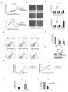
Fig 4. lincRNA-p21 is required for proper apoptotic induction
(A) Increased cell viability of lincRNA-p21 depleted cells. Relative number of siRNA-transfected MEFs treated with 400nM DOX from 24h after transfection (right) or untreated (left) determined by MTT assay. (B) Knockdown of lincRNA-p21 with individual siRNAs increases cell viability. Images of MEFs treated with different individual siRNAs after 48h of DOX treatment (72h post transfection). (C) LincRNA-p21 knockdown doesn't affect cell cycle regulation. Relative cell numbers in each cell cycle phase determined by FACS of BrdU incorporation and PI staining of MEFs treated as in (A). Numbers inside bars represent percentages of cells in each phase. (D) LincRNA-p21 knockdown causes a decrease in cellular apoptosis. p53-reconstituted p53LSL/LSL MEFs transfected with three individual siRNAs targeting lincRNA-p21 (bottom), two independent control siRNAs (top left and middle) or a siRNA pool targeting p53 (top right). 24h after transfection cells were treated with 400nM doxorubicin and 14h later harvested and subjected to FACS analysis. X-axis represents Annexin-V and y-axis 7-AAD staining. The percentage of cells in each quadrant is indicated. (E) Decreased apoptosis caused by lincRNA-p21 knockdown. Percentage of Annexin-V positive cells (FACS) at 38h post transfection (14h of 400nM DOX treatment) in MEFs treated as in (A). (F) LincRNA-p21 knockdown in p53-reconstituted p53LSL/LSL MEFs causes decrease in Caspase 3 cleavage. Levels of cleaved Caspase 3 or control βActin in p53 reconstituted-p53LSL/LSL MEFs treated with the indicated siRNA pools and 500nM DOX for 14h. (G) Decreased cell viability caused by lincRNA-p21 overexpression. Relative numbers of LKR cells overexpressing lincRNA-p21 or control plasmid determined by MTT assay. (H) Overexpression of lincRNA-p21 causes cellular apoptosis under DNA damage induction. Percentage of Annexin-V-positive LKR cells overexpressing lincRNA-p21 or control vector treated with 500nM DOX. (I) LincRNA overexpression doesn't affect cell cycle regulation. Cell cycle analysis of DOX-treated LKR cells overexpressing lincRNA-p21 or control plasmid. All values are the average of 3 biological replicates (+/-STD). *=P< 0.001 relative to controls. Also see Figure S4.
Figure 5
Figure 5. lincRNA-p21 physically interacts with hnRNP-K
(A) Schematic representation of RNA pull-down experiments to identify associated proteins. Biotinylated lincRNA-p21 or antisense RNA were incubated with nuclear extracts, targeted with streptavidin beads, washed, and associated proteins resolved in a gel. Specific bands were cutout and identified by Mass spectrometry. (B) lincRNA-p21 and hnRNP-K specifically interact in vitro. SDS-PAGE gel of proteins bound to lincRNA-p21 (right lane) or antisense RNA (left lane). The highlighted region was submitted for Mass spectrometry identifying hnRNP-K as the band unique to lincRNA-p21. (C) Western blot analysis of the specific association of hnRNP-K with lincRNA-p21. A nonspecific protein (NONO) is shown as control. (D) Association between endogenous lincRNA-p21 and hnRNP-K in the nucleus of DNA damaged MEFs in native conditions. RNA Immunoprecipitation (RIP) enrichment is determined as RNA associated to hnRNP-K IP relative to IgG control. (E) Physical association between lincRNA-p21 and hnRNP-K after chemical crosslinking of life cells. hnRNP-K was immunoprecipitated from nuclear extracts of formaldehyde-crosslinked DNA-damaged MEFs, and associated RNAs detected by RT-qPCR. The relative enrichment is calculated as in (D) and is the median of 3 technical replicates of a representative experiment (+/- STD). (F) lincRNA-p21 binds hnRNP-K through its 5′ terminal region. RNAs corresponding to different fragments of lincRNA-p21 or its antisense sequence (middle and bottom panels) were treated as in (A) and associated hnRNP-K was detected by western blot (top panel). Also see Figure S5. (G) Percentage of Annexin-V positive LKR cells overexpressing the indicated lincRNA-p21 fragments or empty vector as control (average of 3 replicates (+/-STD)). *= P< 0.001.
Figure 6
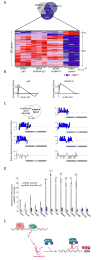
Figure 6. LincRNA-p21 and hnRNP-K corepress genes in the p53 transcriptional response
(A) Many genes are coregulated by p53, LincRNA-p21 and hnRNP-K. Genes affected by knockdown of lincRNA-p21, p53 or hnRNP-K in p53-restored-DNA-damaged p53LSL/LSL MEFs determined by microarray analysis (FDR<0.05). Shades of red or blue represent expression values relative to global median across experiments. Percentages of up- and down-regulated genes are indicated. (B) Genes repressed by lincRNA-p21 are significantly enriched in genes repressed by p53 and hnRNPK. GSEA comparing the genes upregulated on knockdown of LincRNA-p21 and those upregulated upon knockdown of p53 (upper panel) or hnRNP-K (lower panel). The black line represents the observed enrichment score profile of genes in the lincRNA-p21 gene set to the p53 or hnRNP-K gene sets respectively. (C) hnRNP-K associates to promoters of genes corepressed by lincRNA-p21 and p53. Examples of promoters of genes repressed by p53 and lincRNA-p21 (G2e3, Mtap4, Suv39h1 and Vcan) or repressed by lincRNA-p21 but not p53 (Rb1) bound by hnRNP-K (blue) determined by ChIP-chip of hnRNP-K in dox-trated p53-reconstituted p53LSL/LSL MEFs (FDR<0.05). Cdkn2a and Wt1 are negative controls (grey). (D) hnRNP-K binding to lincRNA-p21 and p53 corepressed genes is dependent on lincRNA-p21. Relative enrichment of hnRNP-K (ChIP-qPCR) in the indicated promoter regions in p53-reconstituted p53LSL/LSL MEFs transfected with siRNA lincRNA-p21 or siRNA control and dox-treated determined by ChIP-qPCR (representative of two biological replicates shown +/- STD). (E) Proposed models for the function of licRNA-p21 in the p53 transcriptional response. Induction of p53 activates the transcription of lincRNA-p21 by binding to its promoter (top left). LincRNA-p21 binds to hnRNP-K, and this interaction imparts specificity to genes repressed by p53 induction (top right). See also Figure S6 and Table S3.

"VSports app下载" Comment in

References

    1. Brugarolas J, Chandrasekaran C, Gordon JI, Beach D, Jacks T, Hannon GJ. Radiation-induced cell cycle arrest compromised by p21 deficiency. Nature. 1995;377:552–557. - PubMed
    1. Carninci P. Non-coding RNA transcription: turning on neighbours. Nat Cell Biol. 2008;10:1023–1024. - PubMed
    1. Denisenko ON, Bomsztyk K. The product of the murine homolog of the Drosophila extra sex combs gene displays transcriptional repressor activity. Mol Cell Biol. 1997;17:4707–4717. - PMC - PubMed
    1. el-Deiry WS, Kern SE, Pietenpol JA, Kinzler KW, Vogelstein B. Definition of a consensus binding site for p53. Nat Genet. 1992;1:45–49. - PubMed
    1. Funk WD, Pak DT, Karas RH, Wright WE, Shay JW. A transcriptionally active DNA-binding site for human p53 protein complexes. Mol Cell Biol. 1992;12:2866–2871. - PMC - PubMed

Publication types

Substances

V体育官网入口 - Associated data