Skip to main page content (VSports最新版本)
U.S. flag

An official website of the United States government

Dot gov

The . gov means it’s official. Federal government websites often end in . gov or VSports app下载. mil. Before sharing sensitive information, make sure you’re on a federal government site. .

Https

The site is secure. The https:// ensures that you are connecting to the official website and that any information you provide is encrypted and transmitted securely. V体育官网.

. 2010 Aug 24;103(5):715-26.
doi: 10.1038/sj.bjc.6605823. Epub 2010 Jul 27.

ANO1 amplification and expression in HNSCC with a high propensity for future distant metastasis and its functions in HNSCC cell lines

Affiliations

VSports最新版本 - ANO1 amplification and expression in HNSCC with a high propensity for future distant metastasis and its functions in HNSCC cell lines

C Ayoub et al. Br J Cancer. .

Abstract

Background: Head and neck squamous cell carcinoma (HNSCC) is associated with poor survival. To identify prognostic and diagnostic markers and therapeutic targets, we studied ANO1, a recently identified calcium-activated chloride channel (CaCC) VSports手机版. .

Methods: High-resolution genomic and transcriptomic microarray analysis and functional studies using HNSCC cell line and CaCC inhibitors V体育安卓版. .

Results: Amplification and overexpression of genes within the 11q13 amplicon are associated with the propensity for future distance metastasis of HPV-negative HNSCC. ANO1 was selected for functional studies based on high correlations, cell surface expression and CaCC activity. ANO1 overexpression in cells that express low endogenous levels stimulates cell movement, whereas downregulation in cells with high endogenous levels has the opposite effect. ANO1 overexpression also stimulates attachment, spreading, detachment and invasion, which could account for its effects on migration V体育ios版. CaCC inhibitors decrease movement, suggesting that channel activity is required for the effects of ANO1. In contrast, ANO1 overexpression does not affect cell proliferation. .

Interpretation: ANO1 amplification and expression could be markers for distant metastasis in HNSCC. ANO1 overexpression affects cell properties linked to metastasis. Inhibitors of CaCCs could be used to inhibit the tumourigenic properties of ANO1, whereas activators developed to increase CaCC activity could have adverse effects. VSports最新版本.


"VSports手机版" Figures
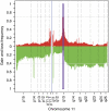
Figure 1
Figure 1
Genomic and transcriptomic microarray analysis of head and neck squamous cell carcinoma (HNSCC) patient samples. Frequency of gains and losses of chromosome 11 sequences plotted along the chromosome. Single-nucleotide polymorphism (SNP) array data (GNL: gain/normal/loss) were used to identify gains (red) and losses (green) along chromosome 11. The boxed area indicates the predominant peak of amplification corresponding to the 11q13 amplicon. The colour reproduction of the figure is available on the html full text version of the paper.
Figure 2
Figure 2
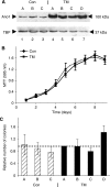
ANO1 overexpression does not affect cell proliferation and growth in soft agar. (A) ANO1 overexpression in stably transfected HEp-2 cells. Extracts of isolated clones (lysis buffer I) were analysed by western blotting using the anti-ANO1 antibody. TM-A, TM-B, TM-C and TM-D: ANO1-overexpressing clones; A, B and C: control clones established using the corresponding empty vector. (B) Cell proliferation measured by the MTT (3-(4,5-Dimethylthiazol-2-yl)-2,5-Diphenyltetrazolium Bromide) assay for the three control (dotted line) and four TM (solid line) clones. MTT conversion was measured at 595 nm. This experiment was repeated three times and similar results were obtained (four independent experiments). (C) Colony formation in soft agar. The number of colonies in each experiment were normalised to TM-A. The Student's t-test for the control clones (hatched) and the ANO1-overexpressing clones (black) gave a P-value of 0.06, which we consider to be not significant. This experiment was repeated once (i.e., two independent experiments) and similar results were obtained.
Figure 3
Figure 3
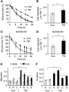
ANO1 overexpression stimulates cell migration and invasion. (A, C) In vitro wound healing assays in the absence (A) or presence of 5 μM aphidicolin (C). The mean values were calculated for the control clones (dotted line) and for the overexpressing clones (solid line). (B, D) Rate of individual cell migration. The distance covered by individual cells was measured at different times of wound healing using Image J. The P-value between the controls (Con) and the overexpressing clones (TM) was ⩽2 × 10−05. (E) Boyden chamber migration assay. The cells that migrated through the porous membranes coated with BSA or collagen I (Col I) were quantified 2.5 h after seeding. The results are the averages from two independent experiments (Student's t-tests: BSA, P=0.35; Col I, P=2 × 10⁻⁴). (F) Boyden chamber invasion assay. Invading cells were quantified 5.5 h after seeding. The results are the averages from two independent experiments (Student's t-test, P=0.002). The dotted lines in (E) and (F) indicate the average values for each group of stable clones. *Indicates statistical significance (P-value <0.05).
Figure 4
Figure 4
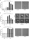
ANO1 overexpression affects cell adhesion, spreading and detachment. (A) ANO1 increases cell adhesion. Left panel: the values are the means of two independent experiments. Student's t-test, Con vs TM, P=2 × 10−04. Right panel: representative photos of cell attachment of control clone A and overexpressing clone TM-D. (B) ANO1 increases cell spreading. Each clone was analysed by time-lapse microscopy at four different positions of the camera. The data represent the proportion of cells that had spread 2 h after plating and correspond to the mean value of three independent experiments. Student's t-test, Con vs TM, P=7 × 10−07. Right panel: representative photos of clone A and clone TM-B cells at different times after plating. (C) TM increases cell detachment. The results are the averages from two independent experiments. Student's t-test, Con vs TM, P=5 × 10⁻¹¹. Right panel: representative photos of cell detachment for clone C and clone TM-D. The dotted lines indicate the average values for the control clones. *Indicates statistical significance (P-value <0.05).
Figure 5
Figure 5
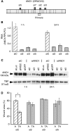
Anoctamin 1 (ANO1) silencing decreases cell migration in the TM-A clone. (A) Schematic representation of the exon structure of ANO1 (ORAOV2), and the localisations of siRNAs 2, 3 and 5, and of the primers used for reverse transcriptase quantitative PCR (RT–qPCR). The primers target all the known ANO1 transcripts. (B) RT–qPCR analysis of ANO1 mRNA expression in clone TM-A transfected with control siRNAs (siC), and siRNAs 2, 3 and 5. At 24 h after transfection, the cells were scrape-wounded, and after a further 1 h and 24 h, RNA was extracted and analysed by RT–qPCR. The siRNA control (SiC) is the average for siLuc, siRISC and siGFP. The data are the mean of two independent experiments. (C) Western blot analysis of ANO1 protein expression in clone TM-A following transfection with the indicated siRNAs, 1 h and 24 after scrape wounding. Cell extracts were prepared with lysis buffer II. (D) Wound closure following siRNA transfection of control clone A and TM-A (TA). Wound areas were measured 12 h after scraping. The graph represents the average of two independent experiments. *Indicates statistical significance (P-value <0.05).
Figure 6
Figure 6
Inhibition of endogenous expression of ANO1 decreases cell migration. (A) Transfection of siRNAs 2, 5 and 7 inhibits ANO1 protein expression in SCC-25 cell line. Cells transfected with siRNAs for 48 h were scrape-wounded. Proteins were extracted 1 and 24 h after cell injury and were analysed by western blotting. (B) ANO1 inhibition decreases migration of SCC-25 cells – shown by in vitro wound healing on SCC-25 transfected with siRNAs (N=2). *Indicates statistical significance (P-value <0.05).
Figure 7
Figure 7
Effect of niflumic acid (NA) on wound closure induced by ANO1. Confluent monolayers of TM-A-, TM-B- and TM-C-overexpressing clones and control clones A, B and C (Con) were wounded with a sterile pipette tip, incubated with NA or solvent alone, and photomicrographs were taken at different times with an inverse microscope (Leica) and the Cool Snap System (Photometrics, Göttingen, Germany). (A) Representative photomicrographs for the clones TM-A and Con-A after 0, 8, 24 and 48 h. (B) Graphical representation of wound area as a function of time and NA concentration. Average values were calculated for three ANO1-overexpressing (TM-A, -B and -C) and three control clones. The difference between the treated (NA in 0.1% dimethylsulphoxide (DMSO)) and untreated (0.1% DMSO) cells was calculated for each group of clones (TM and Con). The results are the average of three independent experiments and two independent woundings for each clone. *Student's t-test, P<0.02.
Figure 8
Figure 8
IC10 (inhibitory concentration of 10%) determination and effects on wound closure of pharmacological inhibitors. (A–D) The Con A–C and TM-A–C clones were seeded at 50 000 cells per well in 96-well plates, incubated with various concentrations of the indicated compounds for 48 h, and then analysed by the MTT assay. The values are the averages for six wells for each of the three clones of a particular type (Con or TM), carried out twice in two experiments. The error bars indicate the s.d. The IC10 values were determined with Matlab software (The Mathworks, Meudon, France). (E) Confluent monolayers of TM clones A, B and C, and Con clones A, B and C were wounded, incubated with the indicated compounds or solvent alone (dimethylsulphoxide (DMSO)), and photomicrographs were taken at different times with an inverse microscope (Leica) and the Snap Cool system. The values shown are for 36 h, four areas for each wound, two wells per clone, averaged over the three Con or TM clones. The wound area is expressed relative to the zero time point. One representative experiment of four is shown. Consistent results were obtained at different time points (24, 36, 48 and 72 h; data not shown). The error bars represent the s.d. The number of stars indicate the P-value exponents from Student's t-tests; **10−2, ***10−3 and ****10−4.

V体育2025版 - References

    1. Akervall JA, Jin Y, Wennerberg JP, Zatterstrom UK, Kjellen E, Mertens F, Willen R, Mandahl N, Heim S, Mitelman F (1995) Chromosomal abnormalities involving 11q13 are associated with poor prognosis in patients with squamous cell carcinoma of the head and neck. Cancer 76: 853–859 - PubMed (V体育2025版)
    1. Akervall JA, Michalides RJ, Mineta H, Balm A, Borg A, Dictor MR, Jin Y, Loftus B, Mertens F, Wennerberg JP (1997) Amplification of cyclin D1 in squamous cell carcinoma of the head and neck and the prognostic value of chromosomal abnormalities and cyclin D1 overexpression. Cancer 79: 380–389 - PubMed
    1. Berenson JR, Koga H, Yang J, Pearl J, Holmes EC, Figlin R (1990) Frequent amplification of the bcl-1 locus in poorly differentiated squamous cell carcinoma of the lung. The Lung Cancer Study Group. Oncogene 5: 1343–1348 - VSports app下载 - PubMed
    1. Beroukhim R, Mermel CH, Porter D, Wei G, Raychaudhuri S, Donovan J, Barretina J, Boehm JS, Dobson J, Urashima M, Mc Henry KT, Pinchback RM, Ligon AH, Cho YJ, Haery L, Greulich H, Reich M, Winckler W, Lawrence MS, Weir BA, Tanaka KE, Chiang DY, Bass AJ, Loo A, Hoffman C, Prensner J, Liefeld T, Gao Q, Yecies D, Signoretti S, Maher E, Kaye FJ, Sasaki H, Tepper JE, Fletcher JA, Tabernero J, Baselga J, Tsao MS, Demichelis F, Rubin MA, Janne PA, Daly MJ, Nucera C, Levine RL, Ebert BL, Gabriel S, Rustgi AK, Antonescu CR, Ladanyi M, Letai A, Garraway LA, Loda M, Beer DG, True LD, Okamoto A, Pomeroy SL, Singer S, Golub TR, Lander ES, Getz G, Sellers WR, Meyerson M (2010) The landscape of somatic copy-number alteration across human cancers. Nature 463: 899–905 - PMC - PubMed
    1. Bignell GR, Greenman CD, Davies H, Butler AP, Edkins S, Andrews JM, Buck G, Chen L, Beare D, Latimer C, Widaa S, Hinton J, Fahey C, Fu B, Swamy S, Dalgliesh GL, Tch BT, Deloukas P, Yang F, Campbell PJ, Futreal PA, Stratton MR (2010) Signatures of mutation and selection in the cancer genome. Nature 463: 893–898 - PMC - PubMed

Publication types

"VSports" MeSH terms