Skip to main page content
U.S. flag

An official website of the United States government

Dot gov

The . gov means it’s official. Federal government websites often end in VSports app下载. gov or . mil. Before sharing sensitive information, make sure you’re on a federal government site. .

Https

The site is secure. The https:// ensures that you are connecting to the official website and that any information you provide is encrypted and transmitted securely. V体育官网.

Review
. 2010 Jun 26;3(6):557-69.

Enchondromatosis: insights on the different subtypes

Affiliations
Review

Enchondromatosis: insights on the different subtypes

Twinkal C Pansuriya et al. Int J Clin Exp Pathol. .

Abstract

Enchondromatosis is a rare, heterogeneous skeletal disorder in which patients have multiple enchondromas. Enchondromas are benign hyaline cartilage forming tumors in the medulla of metaphyseal bone. The disorder manifests itself early in childhood without any significant gender bias. Enchondromatosis encompasses several different subtypes of which Ollier disease and Maffucci syndrome are most common, while the other subtypes (metachondromatosis, genochondromatosis, spondyloenchondrodysplasia, dysspondyloenchondromatosis and cheirospondyloenchondromatosis) are extremely rare. Most subtypes are non-hereditary, while some are autosomal dominant or recessive. The gene(s) causing the different enchondromatosis syndromes are largely unknown. They should be distinguished and adequately diagnosed, not only to guide therapeutic decisions and genetic counseling, but also with respect to research into their etiology. For a longtime enchondromas have been considered a developmental disorder caused by the failure of normal endochondral bone formation. With the identification of genetic abnormalities in enchondromas however, they were being thought of as neoplasms. Active hedgehog signaling is reported to be important for enchondroma development and PTH1R mutations have been identified in approximately 10% of Ollier patients VSports手机版. One can therefore speculate that the gene(s) causing the different enchondromatosis subtypes are involved in hedgehog/PTH1R growth plate signaling. Adequate distinction within future studies will shed light on whether these subtypes are different ends of a spectrum caused by a single gene, or that they represent truely different diseases. We therefore review the available clinical information for all enchondromatosis subtypes and discuss the little molecular data available hinting towards their cause. .

Keywords: Maffucci syndrome; Ollier disease; central chondrosarcoma; enchondroma; enchondromatosis; metachondromatosis V体育安卓版. .


Figures

Figure 1
Figure 1
Enchondromatosis sub-types. Classification diagram for patients with multiple enchondromas based on spinal involvement and genetic transmission.
Figure 2
Figure 2
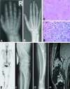
Ollier disease. (A) 4-year-old female patient with Ollier disease. Multiple enchondromas, manifesting as central end eccentric osteolytic lesions and deformities in the metacarpals and phalanges of the fourth and fifth ray of the right hand. (B) Same patient as in Fig. 2A 13 years later. The enchondromas have increased in size and some are more evidently visible compared to the previous study. This has resulted in deformity of the fourth finger. (C) Histology of enchondroma of long bone from Ollier patient showing moderate cellularity and abundance of hyaline cartilage matrix (200 times magnification). (D) Histology of chondrosarcoma grade II of the long bone from Ollier patient showing increased cellularity and atypical chondrocytes (200 times magnification). (E) Technetium-99m bone scintigraphy, anterior projection, demonstrates shortening of the right lower limb. Varus deformity of the femur and valgus deformity of the tibia. Multiple areas of focally increased uptake of the tracer in femur and tibia. (F-G) Anteroposterior radiograph of the right knee and lower leg of same patient as 2E. Deformity of both the distal femur and the tibia and fibula. Structural changes in the marrow cavity and cortical bone of femur and tibia consisting of osteolysis and osteo-sclerosis. Specifically in the proximal tibia areas with mineralization in the sense of calcifications can be appreciated (arrow). The appearance is consistent with multiple chondromatous tumors. (H) Coronal fat-suppressed T1-weighted magnetic resonance image after intravenous contrast administration of the femur. Varus deformation of the femur. Multiple, partially lobulated, areas with increased signal intensity due to enhancement of the chondromatous lesions. Large lesion in the distal diaphysis of the femur, of which histology showed a chondrosarcoma (arrow). The enhancement demonstrates rings and arcs (also known as septal or nodular enhancement) consistent with the chondromatous nature of the lesions.
Figure 3
Figure 3
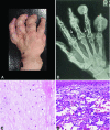
Maffucci Syndrome: (A) Hand of a patient with Maffucci syndrome showing deformities due to multiple enchondromas and a superficial haemangioma. (B) Radiograph of a patient with Maffucci syndrome. Multiple enchondromas with and without soft tissue extension in the second to fifth digit and the fifth metacarpal bone. In addition phleboliths in the soft tissue at the basis of the second and fourth finger (arrows) indicating haemangiomas. (C) Histology of enchondroma and (D) Haemangioma (400 times magnification).
Figure 4
Figure 4
Metachondromatosis. (A) Radiograph of the pelvis with an enchondroma in the right proximal femur (arrow) adjacent to the apophysis of the greater trochanter; (B) radiograph of both hands showing multiple osteochondromas pointing towards the epiphyses. Enchondroma in the proximal phalanx of the left third digit (arrow); (C, E) micrographs of osteochondroma-like lesions (D) magnification of C. These lesions are histologically indistinguishable from conventional osteochondroma recapitulating the normal growth plate architecture (reproduced with permission from [64]).

References

    1. Lucas DR, Bridge JA. Chondromas: enchondroma, periosteal chondroma,and enchondro matosis. World Health Organization classification of tumours. In: Fletcher CDM, Unni KK, Mertens F, editors. Pathology and genetics of tumours of soft tissue and bone. Lyon: IARC Press; 2002. pp. 237–40.
    1. Silve C, Juppner H. Ollier disease. Orphanet J Rare Dis. 2006;1:37. - PMC (VSports在线直播) - PubMed
    1. Spranger JW, Brill PW, Poznanski AK. second ed. New York: Oxford University Press; 2002. Bone Dysplasias, An Atlas of Genetic Disorders of Skeletal Development; pp. 554–70.
    1. Unni KK. Cartilaginous lesions of bone. J Orthop Sci. 2001;6:457–72. - "VSports在线直播" PubMed
    1. Loder RT, Sundberg S, Gabriel K, Mehbod A, Meyer C. Determination of bone age in children with cartilaginous dysplasia (multiple hereditary osteochondromatosis and Ollier's enchondromatosis) J Pediatr Orthop. 2004;24:102–8. - PubMed

Publication types

MeSH terms