"VSports app下载" Skip to main page content
U.S. flag

An official website of the United States government

Dot gov

The . gov means it’s official VSports app下载. Federal government websites often end in . gov or . mil. Before sharing sensitive information, make sure you’re on a federal government site. .

Https

The site is secure V体育官网. The https:// ensures that you are connecting to the official website and that any information you provide is encrypted and transmitted securely. .

. 2010 Jul 26;190(2):233-45.
doi: 10.1083/jcb.201001129.

Ras-mediated activation of the TORC2-PKB pathway is critical for chemotaxis

Affiliations

"V体育2025版" Ras-mediated activation of the TORC2-PKB pathway is critical for chemotaxis

Huaqing Cai (V体育安卓版) et al. J Cell Biol. .

Abstract

In chemotactic cells, G protein-coupled receptors activate Ras proteins, but it is unclear how Ras-associated pathways link extracellular signaling to cell migration VSports手机版. We show that, in Dictyostelium discoideum, activated forms of RasC prolong the time course of TORC2 (target of rapamycin [Tor] complex 2)-mediated activation of a myristoylated protein kinase B (PKB; PKBR1) and the phosphorylation of PKB substrates, independently of phosphatidylinositol-(3,4,5)-trisphosphate. Paralleling these changes, the kinetics of chemoattractant-induced adenylyl cyclase activation and actin polymerization are extended, pseudopodial activity is increased and mislocalized, and chemotaxis is impaired. The effects of activated RasC are suppressed by deletion of the TORC2 subunit PiaA. In vitro RasC(Q62L)-dependent PKB phosphorylation can be rapidly initiated by the addition of a PiaA-associated immunocomplex to membranes of TORC2-deficient cells and blocked by TOR-specific inhibitor PP242. Furthermore, TORC2 binds specifically to the activated form of RasC. These results demonstrate that RasC is an upstream regulator of TORC2 and that the TORC2-PKB signaling mediates effects of activated Ras proteins on the cytoskeleton and cell migration. .


Figures

Figure 1.
Figure 1.
RasC is required for the activation of the PKB pathway. (A) Schematic representation of the activation of PKBR1 and PKBA. PKBR1 is tethered to the plasma membrane via myristoylation, whereas PKBA is recruited to the plasma membrane by PIP3 via the PH domain. Upon chemoattractant stimulation, the two PKBs are activated through phosphorylation of their HMs by TORC2 and ALs by PDK. (B) Wild-type (WT) or rasC cells were stimulated with cAMP, sampled at the indicated time points, and probed with anti–phospho-HM (first panel), anti–phospho-AL (second panel), or anti–phospho-substrate antibodies (third panel). The protein-transferred membrane was stained with Coomassie brilliant blue (CBB) and shown as loading control (forth panel). Seven proteins, including TalinB, GEFN, GEFS, PI5K, and GacQ, are confirmed PKB substrates. Note that TalinB co-migrates with a non-PKB substrate band, and unmarked bands at the bottom part of the third panel are not PKB substrates (Kamimura et al., 2008). (C) Quantitative densitometry of the first and second panels of B, showing the mean intensity ± SD of the respective bands from three independent experiments. (D) Densitometric scan of the 60-s lanes of the third panel of B (wild type, black line; rasC, gray line).
Figure 2.
Figure 2.
Persistently activated RasC alters the time course of PKBR1 and PKB substrate phosphorylation. (A) Flag-RasC or -RasCQ62L was expressed under the control of a doxycycline-inducible promoter in rasC cells. Cells developed in the presence or absence of doxycycline were stimulated with cAMP, sampled at the indicated time points, and probed with phospho-specific antibodies or anti-Flag antibody. The protein-transferred membrane was stained with Coomassie brilliant blue (CBB) and shown as loading control. (B) Quantitative densitometry of the first and second panels of A, showing the mean intensity ± SD of the respective bands from four independent experiments. (C) Samples were probed with anti–phospho-substrate antibody (top). Densitometric scans of the 240-s lanes are shown in bottom panel (Flag-RasC, gray line; Flag-RasCQ62L, black line).
Figure 3.
Figure 3.
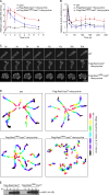
Chemotactic responses are altered in cells with prolonged PKBR1 signaling. (A) ACA activation upon uniform cAMP stimulation was measured in rasC cells or rasC cells induced to express Flag-RasC or -RasCQ62L. The data represent the range (mean ± standard error) of two independent experiments. The basal activities ranged from 1.9 pmol/min/mg protein for rasC to 2.5 pmol/min/mg protein for Flag-RasC/rasC and 3.8 pmol/min/mg protein for Flag-RasCQ62L/rasC cells. (B) Actin polymerization was measured in wild-type (WT) cells or rasC cells expressing Flag-RasC or -RasCQ62L. Cells were stimulated with cAMP and lysed with Triton X-100 buffer. The Triton-insoluble pellet fraction was analyzed by SDS-PAGE and Coomassie blue staining. The amount of actin in the pellet was quantified by densitometry. The data represent mean ± SD of three independent experiments. A.U., arbitrary unit. (C) The translocation of LimEΔcoil-RFP was recorded by fluorescence microscopy with images taken every 3 s. Frames from the indicated time points after the addition of cAMP are shown. (D) The chemotactic movements to a micropipette releasing 1 µM cAMP (black dot) were recorded by time-lapse microscopy. Images from frames at 2.5-min intervals were processed to outline the cells, color coded for each time point, and overlaid. (E) The expression of cAR1 is comparable in rasC cells induced to express Flag-RasC or -RasCQ62L. At the indicated time points after initiation of development, CHAPS-insoluble membrane fractions were prepared as described previously (Xiao et al., 1997) and probed with anti-cAR1 antibody. Bar, 10 µm.
Figure 4.
Figure 4.
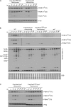
Deletion of the TORC2 component PiaA suppresses the prolonged phosphorylation of PKBR1 and PKB substrates caused by RasCQ62L. (A–C) Flag-RasC or -RasCQ62L was expressed under the control of a doxycycline-inducible promoter in wild-type (WT; A), piaA (B), or aca cells (C). Cells were stimulated with cAMP, sampled at the indicated time points, and probed with phospho-specific antibodies or anti-Flag antibody. The protein-transferred membrane was stained with Coomassie brilliant blue (CBB) and shown as loading control. Vertical black lines indicate that intervening lanes have been spliced out.
Figure 5.
Figure 5.
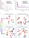
Deletion of PiaA suppresses the unregulated chemotactic responses caused by RasCQ62L. (A and B) ACA activation and actin polymerization were measured in wild-type (WT) or piaA cells induced to express Flag-RasC or -RasCQ62L. ACA data represent the range (mean ± standard error) of two independent experiments. The basal activities ranged from 15.2 pmol/min/mg protein for Flag-RasC/piaA and -RasCQ62L/piaA cells to 17.9 pmol/min/mg protein for Flag-RasC/WT and 19.2 pmol/min/mg protein for Flag-RasCQ62L/WT cells. For the actin polymerization assay, the data represent mean ± SD of three independent experiments. A.U., arbitrary unit. (C) The micropipette assay was performed in aca or piaA cells induced to express Flag-RasC or -RasCQ62L.
Figure 6.
Figure 6.
PIP3 is not required for RasC-mediated activation of the TORC2–PKBR1 pathway. (A) PHcrac-GFP translocation assay, which measures PIP3 content on the membrane, was performed in rasC cells induced to express Flag-RasC or -RasCQ62L. (B) Phosphorylation of PKB was measured in wild-type (WT), pi3k1/2, and pi3k1/2 cells expressing Flag-RasC or -RasCQ62L. The protein-transferred membrane was stained with Coomassie brilliant blue (CBB) and shown as loading control. (C) Phosphorylation of PKB was measured in wild-type and pten cells. The protein-transferred membrane was stained with Coomassie brilliant blue and shown as loading control. (B and C) Vertical black lines indicate that intervening lanes have been spliced out.
Figure 7.
Figure 7.
RasCQ62L activates TORC2-mediated phosphorylation of PKB in vitro. (A) Wild-type cell lysates were stimulated with GTP-γS alone or in the presence of GST or GST-RBD and then probed with phospho-specific antibodies. The protein-transferred membrane was stained with Coomassie brilliant blue (CBB) and shown as loading control. (B) The indicated cell lines were mixed, lysed, incubated in the presence or absence of GST or GST-RBD for 12 min, and then probed with phospho-specific antibodies. The protein-transferred membrane was stained with Coomassie brilliant blue and shown as loading control. Cells were not stimulated with chemoattractant, nor was GTP-γS added to the reaction. WT, wild type. (C) Quantitative densitometry of the time courses of reactions 3, 4, and 5 of B. The data represent mean ± SD of three independent experiments. A.U., arbitrary unit. (D) piaA cells expressing Flag-RasCQ62L were mixed with wild-type cells in the presence of increasing concentrations of GDP-βS, filter lysed, incubated on ice for 12 min, and then probed with anti–phospho-HM antibody.
Figure 8.
Figure 8.
Reconstitution of PKB activation in vitro with immunopurified TORC2. (A) Cell lysates from piaA cells expressing Flag-RasC or -RasCQ62L were mixed with HSS made from wild-type (WT) cells or piaA cells expressing Flag-PiaA or with Flag-eluted immunoprecipitate from Flag/piaA cells or Flag-PiaA/piaA cells. (B) Membrane fractions (MF) prepared from Flag-RasC/piaA or Flag-RasCQ62L /piaA cells were mixed with Flag-eluted immunoprecipitate from Flag/piaA cells or Flag-PiaA/piaA cells. (C) Cell lysates from rip3 cells expressing Flag-RasC or -RasCQ62L were mixed with Flag-eluted immunoprecipitate from Flag/piaA cells or Flag-PiaA/piaA cells. (D) Cell lysates from piaA cells expressing Flag-RasCQ62L were mixed with Flag-eluted immunoprecipitate from Flag-PiaA/piaA cells or Flag-PiaA/rip3 cells. (E) Flag-eluted immunoprecipitate from Flag-PiaA/piaA cells was treated with increasing concentrations of PP242 before being mixed with cell lysates from piaA cells expressing Flag-RasCQ62L. (F) Whole cell extracts were prepared from rip3 cells expressing T7-Rip3 and Flag-RasC, T7-Rip3 and Flag-RasCQ62L, or Flag-RasCQ62L alone. Immunoprecipitation (IP) was performed using anti-T7 antibody, and samples were immunoblotted (IB) with anti-T7 or anti-Flag antibodies.
Figure 9.
Figure 9.
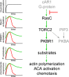
Schematic diagram of RasC-mediated signaling pathways that control chemotaxis. The chemoattractant cAMP signals through the G protein–coupled receptor cAR1 to RasC, leading to TORC2-mediated activation of PKBR1 and PKBA. The activation of PKBA also depends on recruitment to PIP3. Together, the two PKBs phosphorylate a series of substrates and play a critical role in ACA activation, actin polymerization, and chemotaxis. The graphs to the left schematically illustrate the kinetics of G protein α and βγ subunits dissociation, RasC activation, PKB phosphorylation, PKB substrate phosphorylation, and chemotactic responses. The red line shows the typical wild-type responses that are rapidly shut off during persistent stimulation. The green line shows the responses in cells in which the inhibitory signal indicated by (⊥) is bypassed by RasCQ62L.

VSports app下载 - References

    1. Cantley L.C. 2002. The phosphoinositide 3-kinase pathway. Science. 296:1655–1657 10.1126/science.296.5573.1655 - DOI (VSports手机版) - PubMed
    1. Chen M.Y., Long Y., Devreotes P.N. 1997. A novel cytosolic regulator, Pianissimo, is required for chemoattractant receptor and G protein-mediated activation of the 12 transmembrane domain adenylyl cyclase in Dictyostelium. Genes Dev. 11:3218–3231 10.1101/gad.11.23.3218 - DOI - PMC - PubMed
    1. Faix J., Kreppel L., Shaulsky G., Schleicher M., Kimmel A.R. 2004. A rapid and efficient method to generate multiple gene disruptions in Dictyostelium discoideum using a single selectable marker and the Cre-loxP system. Nucleic Acids Res. 32:e143 10.1093/nar/gnh136 - DOI - PMC - PubMed
    1. Feig L.A., Cooper G.M. 1988. Relationship among guanine nucleotide exchange, GTP hydrolysis, and transforming potential of mutated ras proteins. Mol. Cell. Biol. 8:2472–2478 - PMC (VSports最新版本) - PubMed
    1. Feldman M.E., Apsel B., Uotila A., Loewith R., Knight Z.A., Ruggero D., Shokat K.M. 2009. Active-site inhibitors of mTOR target rapamycin-resistant outputs of mTORC1 and mTORC2. PLoS Biol. 7:e38 10.1371/journal.pbio.1000038 - DOI - PMC - PubMed

"VSports app下载" Publication types

MeSH terms

LinkOut - more resources