Skip to main page content
U.S. flag

An official website of the United States government

Dot gov

The . gov means it’s official VSports app下载. Federal government websites often end in . gov or . mil. Before sharing sensitive information, make sure you’re on a federal government site. .

Https

The site is secure. The https:// ensures that you are connecting to the official website and that any information you provide is encrypted and transmitted securely. V体育官网.

. 2010 Sep 10;285(37):28715-22.
doi: 10.1074/jbc.M110.133355. Epub 2010 Jun 30.

"VSports" Autophagy facilitates IFN-gamma-induced Jak2-STAT1 activation and cellular inflammation

Affiliations

Autophagy facilitates IFN-gamma-induced Jak2-STAT1 activation and cellular inflammation

Yu-Ping Chang et al. J Biol Chem. .

Abstract

Autophagy is regulated for IFN-gamma-mediated antimicrobial efficacy; however, its molecular effects for IFN-gamma signaling are largely unknown. Here, we show that autophagy facilitates IFN-gamma-activated Jak2-STAT1. IFN-gamma induces autophagy in wild-type but not in autophagy protein 5 (Atg5(-/-))-deficient mouse embryonic fibroblasts (MEFs), and, autophagy-dependently, IFN-gamma induces IFN regulatory factor 1 and cellular inflammatory responses. Pharmacologically inhibiting autophagy using 3-methyladenine, a known inhibitor of class III phosphatidylinositol 3-kinase, confirms these effects VSports手机版. Either Atg5(-/-) or Atg7(-/-) MEFs are, independent of changes in IFN-gamma receptor expression, resistant to IFN-gamma-activated Jak2-STAT1, which suggests that autophagy is important for IFN-gamma signal transduction. Lentivirus-based short hairpin RNA for Atg5 knockdown confirmed the importance of autophagy for IFN-gamma-activated STAT1. Without autophagy, reactive oxygen species increase and cause SHP2 (Src homology-2 domain-containing phosphatase 2)-regulated STAT1 inactivation. Inhibiting SHP2 reversed both cellular inflammation and the IFN-gamma-induced activation of STAT1 in Atg5(-/-) MEFs. Our study provides evidence that there is a link between autophagy and both IFN-gamma signaling and cellular inflammation and that autophagy, because it inhibits the expression of reactive oxygen species and SHP2, is pivotal for Jak2-STAT1 activation. .


Figures
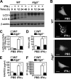
FIGURE 1.
FIGURE 1.
IFN-γ-induced autophagy was critical for cellular inflammation. A, Western blotting was used to determine LC3 conversion in IFN-γ (10 ng/ml)-treated WT and Atg5−/− MEFs. β-Actin was the internal control. Data are representative of three individual experiments. B, confocal fluorescence microscopic observation of EGFP-LC3 punctate formation 6 h after IFN-γ (10 ng/ml) treatment in WT and Atg5−/− MEFs is shown. Scale bars, 20 μm. C, luciferase reporter assay of IRF1 promoter transactivation 1 h after IFN-γ (10 ng/ml) treatment in WT and Atg5−/− MEFs is shown. D, Griess reagent was used to detect the generation of nitrite 48 h after IFN-γ (10 ng/ml) treatment. E and F, ELISA was used to measure RANTES and IP-10 production 24 h after IFN-γ (10 ng/ml) treatment. Data, obtained from triplicate cultures, are means ± S.D. (error bars). *, p < 0.05.
FIGURE 2.
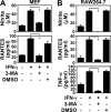
FIGURE 2.
Inhibiting autophagy reduced IFN-γ-induced inflammation. Griess reagent was used to detect nitrite generation 48 h after IFN-γ (10 ng/ml) treatment in WT MEFs (A) and RAW264.7 cells (B) pretreated for 0.5 h with (+) and without (−) 3-MA (1 mm). ELISA was used to measure RANTES and TNF-α 24 h after treatment. Dimethyl sulfoxide (DMSO) was the control. Data, obtained from triplicate cultures, are means ± S.D. (error bars). *, p < 0.05.
FIGURE 3.
FIGURE 3.
Effects of autophagy on the expression of IFN-γ receptors and the activation of Jak2 signaling. A, flow cytometry was used to detect the expression of IFNGR1 and IFNGR2 using specific antibodies in WT and Atg5−/− MEFs. The percentages of positive cells are shown. B, after 0.25 h of IFN-γ (10 ng/ml) treatment, co-immunoprecipitation (IP) was used to detect Jak2 interaction with IFNGR2. Western blotting (IB) was used to determine the expression of Jak2 and IFNGR2. H, heavy chain; L, light chain; NB, nonspecific binding. C, after IFN-γ (10 ng/ml) treatment, Western blotting was used to determine the time kinetic phosphorylation of Jak2 (Tyr1007/Tyr1008) and Jak1 (Tyr1022/Tyr1023). β-Actin was the internal control. Data are representative of three individual experiments.
FIGURE 4.
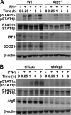
FIGURE 4.
Autophagy was important for IFN-γ-activated STAT1. A, after IFN-γ (10 ng/ml) treatment, Western blotting was used to determine the time kinetic phosphorylation of STAT1α/β (Tyr701) as well as IRF1 and SOCS1 expression in WT and Atg5−/− MEFs. B, HMEC-1 cells were pretreated with or without lentivirus-based short hairpin Atg5 RNA (shAtg5) or control luciferase-shRNA (shLuc) transfection and then treated with IFN-γ (10 ng/ml) for the indicated times. Western blotting was used to determine the expression of phosphorylation of STAT1α/β (Tyr701) and Atg5. β-Actin was the internal control. Data are representative of three individual experiments.
FIGURE 5.
FIGURE 5.
Autophagy was required for IFN-γ-activated Jak2-STAT1 and inflammation. Western blotting was used to determine LC3 conversion (A) and the time kinetic phosphorylation of Jak2 (Tyr1007/Tyr1008) and STAT1α/β (Tyr701) (B) in IFN-γ (10 ng/ml)-treated WT and Atg7−/− MEFs. β-Actin was the internal control. Data are representative of three individual experiments. C, Griess reagent was used to detect nitrite generation 48 h after IFN-γ (10 ng/ml) treatment. D, ELISA was used to measure IP-10 production 24 h after IFN-γ (10 ng/ml) treatment. Data, obtained from triplicate cultures, are means ± S.D. (error bars). *, p < 0.05.
FIGURE 6.
FIGURE 6.
ROS generation was deregulated in the absence of autophagy and negatively regulated IFN-γ-induced STAT1 activation. A, confocal fluorescence microscopic observation of the co-localization (Merge, yellow) of EGFP-LC3 punctate formation (green) and MitoSOX Red staining (red) in WT MEFs with or without IFN-γ (10 ng/ml) treatment for 3 h. 3D, three-dimensional. Scale bars, 20 μm. B, Western blotting, PCR, and flow cytometry, with COX IV antibodies, COX I primer, and MitoTracker Green staining, respectively, were used to detect mitochondria expression in WT and Atg5−/− MEFs. The ratios of mitochondrial DNA COX I to the total genomic DNA GAPDH were calculated based on the intensities of their PCR product in agarose gel. MFI, mean fluorescence intensity. C, CM-H2DCFDA staining and a microplate reader were used to measure ROS generation. Data, obtained from triplicate cultures, are means ± S.D. (error bars). *, p < 0.05. D, Western blotting was used to determine IFN-γ (10 ng/ml)-induced phosphorylation of STAT1α/β (Tyr701) in WT MEFs 0.25 h after they had been pretreated with (+) and without (−) caffeic acid phenethyl ester (CAPE, 25 μm) or H2O2 (10 mm) for 0.5 h. β-Actin was the internal control. Data are representative of three individual experiments. E, with (+) and without (−) H2O2 pretreatment for 0.5 h, generation of nitrite in IFN-γ (10 ng/ml)-treated WT MEFs was detected using Griess reagent at 48 h. Data, obtained from triplicate cultures, are means ± S.D. (error bars). *, p < 0.05.
FIGURE 7.
FIGURE 7.
ROS-regulated SHP2 inhibited IFN-γ-activated STAT1 in the absence of autophagy. A, Western blotting was used to determine IFN-γ (10 ng/ml)-induced phosphorylation of STAT1α/β (Tyr701) in WT MEFs 0.25 h after they had been pretreated with (+) and without (−) SHP2-shRNA clone 2 (shSHP2–2) or control luciferase-shRNA (shLuc) transfection and then with H2O2 (10 mm) for 0.5 h. B, after IFN-γ (10 ng/ml) treatment with (+) and without (−) SHP2-shRNAs (shSHP2) or control (shLuc) transfection, Western blotting was used to determine the phosphorylation of STAT1α/β (Tyr701) in Atg5−/− MEFs. shSHP2-1, inefficiently silenced; shSHP2-2, efficiently silenced. β-Actin was the internal control. Data are representative of three individual experiments.

References

    1. Delgado M. A., Elmaoued R. A., Davis A. S., Kyei G., Deretic V. (2008) EMBO J. 27, 1110–1121 - PMC (VSports在线直播) - PubMed
    1. Deretic V. (2009) Curr. Opin. Immunol. 21, 53–62 - PMC - PubMed
    1. Codogno P., Meijer A. J. (2005) Cell Death Differ. 12, (Suppl. 2), 1509–1518 - PubMed
    1. Lee H. K., Lund J. M., Ramanathan B., Mizushima N., Iwasaki A. (2007) Science 315, 1398–1401 - PubMed
    1. English L., Chemali M., Duron J., Rondeau C., Laplante A., Gingras D., Alexander D., Leib D., Norbury C., Lippé R., Desjardins M. (2009) Nat. Immunol. 10, 480–487 - PMC (VSports最新版本) - PubMed

Publication types

MeSH terms

Substances