Skip to main page content
U.S. flag

An official website of the United States government

Dot gov

The . gov means it’s official. Federal government websites often end in VSports app下载. gov or . mil. Before sharing sensitive information, make sure you’re on a federal government site. .

Https

The site is secure. The https:// ensures that you are connecting to the official website and that any information you provide is encrypted and transmitted securely V体育官网. .

. 2010 Nov;69(11):2042-50.
doi: 10.1136/ard.2009.126383. Epub 2010 Jun 15.

Generation and characterisation of therapeutic tolerogenic dendritic cells for rheumatoid arthritis

Affiliations

Generation and characterisation of therapeutic tolerogenic dendritic cells for rheumatoid arthritis

Rachel A Harry et al. Ann Rheum Dis. 2010 Nov.

Abstract

Objectives: Tolerogenic dendritic cells (tolDCs) constitute a promising experimental treatment for targeting autoreactive T cells in autoimmune diseases, including rheumatoid arthritis (RA). The authors' goal is to bring tolDC therapy for RA to the clinic VSports手机版. Here the authors address key translational issues related to the manufacturing of tolDCs from RA patients with current good manufacturing practice (cGMP)-compliant reagents, the stability of tolDCs, and the selection of suitable quality control markers. .

Methods: Human monocyte-derived tolDCs were established from RA patients and healthy controls (HCs) using the immunosuppressive drugs dexamethasone and vitamin D₃, and the cGMP-grade immunomodulator, monophosphoryl lipid A, in the cGMP-compliant medium, CellGroDC V体育安卓版. The functionality of tolDCs and tolDC-modulated autologous CD4 T cells was determined by flow cytometry, [³H]thymidine incorporation and ELISA. .

Results: Clinical-grade tolDCs established from patients with RA exhibit a typical tolerogenic phenotype of reduced costimulatory molecules, low production of proinflammatory cytokines and impaired stimulation of autologous antigen-specific T cells, comparable to HC tolDCs. Toll-like receptor 2 (TLR-2) was highly expressed by tolDCs but not mature DCs V体育ios版. Furthermore, tolDCs suppressed mature DC-induced T cell proliferation, interferon γ and interleukin 17 production, and rendered T cells hyporesponsive to further stimulation. Importantly, tolDCs were phenotypically stable in the absence of immunosuppressive drugs and were refractory to further challenge with proinflammatory mediators. .

Conclusions: tolDCs established from patients with RA are comparable to those derived from healthy donors. TLR-2 was identified as an ideal marker for quality control of tolDCs VSports最新版本. Potently tolerogenic and highly stable, these tolDCs are a promising cellular therapeutic for tailored immunomodulation in the treatment of RA. .


Conflict of interest statement

Competing interests None.

Figures

Figure 1
Figure 1
Rheumatoid arthritis (RA)-derived tolerogenic dendritic cells (tolDCs) exhibit a semimature phenotype and are comparable to healthy control (HC) tolDCs. (A) HC- and RA-derived DC expression of maturation-associated markers of immature DCs (iDCs), mature DCs (matDCs) and tolDCs. Histograms are representative of five independent donors. (B) Mean expression of HC and RA patient tolDCs and matDCs is shown relative to iDCs. Expression of each marker (geometric mean fluorescence intensity (GMFI)) was normalised relative to expression by iDCs (100%) for each independent donor. Phenotypic analysis of maturation-associated markers was determined 24 h after maturation with 1 µg/ml monophosphoryl lipid A in the presence or absence of dexamethasone and vitamin D3. Data are expressed as the mean±SEM for five independent donors. Mean GMFI for HC iDCs vs RA iDCs are: MHC II, 949.8±161.3 vs 1110±178.3; CD40, 19.8±7.4 vs 20.4±6.6; CD80, 12.3±1.7 vs 11.2±1.4; CD83, 7.9±0.1 vs 9.4±1.2; CD86, 11.5±2.5 vs 21.6±5.8; toll-like receptor 2 (TLR-2), 89.9±11.1 vs 76.2±4.2. No significant difference was detected between RA and HC DC populations. *p≤0.05, **p≤0.01, ***p≤0.001 (analysis of variance). MHC, major histocompatibility complex; TLR-2, toll-like receptor-2.
Figure 2
Figure 2
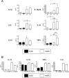
Healthy control (HC) and rheumatoid arthritis (RA) tolerogenic dendritic cells (tolDCs) have an anti-inflammatory phenotpye. (A) Mean cytokine production by tolDCs and mature DCs (matDCs) in response to CD40 activation. At 24 h after maturation with 1 µg/ml monophosphoryl lipid A (MPLA) in the presence or absence of dexamethasone (Dex) and vitamin D3 (VitD3), DCs were washed, and 105 tolDCs or matDCs cocultured at a 1:1 ratio with the J588L-CD40L-expressing cell line in the absence of Dex and VitD3 for a further 24 h. Supernatants were harvested for cytokine analysis by ELISA. Data are expressed as the mean±SEM for at least four HCs and seven RA-independent donors. (B) Representative RA donor DC-derived cytokine production when unloaded (UL) or loaded with 10 µg/ml human cartilage glycoprotein 39 (HCgp39) as a model antigen, during maturation with 1 µg/ml MPLA. Unloaded and antigen-loaded DCs were activated via CD40 as described in (A). Data are representative of four independent donors. Cytokine production was determined by ELISA.
Figure 3
Figure 3
Antigen-specific T cells exhibit a reduced responsiveness to tolerogenic dendritic cells (tolDCs). (A) 105 healthy control (HC) and RA CD4 T cells were stimulated with 104 autologous unloaded or purified protein derivative (PPD)-loaded (1 µg/ml) tolDCs or mature DCs (matDCs), and proliferation determined at various time points. Data are expressed as the mean±SEM of triplicate wells, representative of three independent experiments. (B) Mean healthy control (HC) CD4 T cell proliferation and cytokine production as a percentage of matDC-induced T cell responses. HC CD4 T cells (105) were stimulated at a 10:1 ratio with 104 PPD-loaded tolDCs or matDCs. Cytokine production and proliferation were determined on day 6 and 7, respectively. Data are expressed as the mean of three independent experiments. Mean values for matDC-induced proliferation were 33 642±18807 cpm, interferon (IFN)γ was 16.9±8.1 ng/ml and interleukin (IL)-17 was 359.5±328.8 pg/ml. T cell proliferation was determined by [3H]thymidine incorporation, and cytokine production was quantitated by ELISA. *p≤0.05, **p≤0.01, ***p≤0.001 as compared with matDCs (two-way analysis of variance (A) and Student t test (B)).
Figure 4
Figure 4
Tolerogenic dendritic cells (tolDCs) induce T cell hyporesponsiveness. (A) 105 CD4 T cells were primed with the indicated numbers of toxic shock syndrome toxin 1 (TSST-1)-loaded (5 ng/ml) autologous tolDCs or mature DCs (matDCs) and proliferation of carboxyfluorescein succinimidyl ester (CFSE+) T cell receptor β-chain variable region 2 (TCR-Vβ2)-stained cells quantitated at day 5. Values represent percentage of cells as a percentage of CD4 T cell population. Percentage of Vβ2+ cells in starting CD4 population was 8.9±0.95%. (B) and (C) T cells primed with TSST-1-loaded (5 ng/ml) tolDCs (Ttol) or matDCs (Tmat) (10:1 ratio) for 6 days were rested with interleukin (IL)-2 (20 IU/ml) for 4 days before CFSE labelling and restimulation at a 10:1 ratio with TSST-1-loaded matDCs on day 10. (B) Day 13 T cell proliferation was determined by CFSE dilution. Values represent the percentage of proliferating cells. (C) Day 13 intracellular IL-17 and interferon (IFN)γ production was quantitated after 5 h stimulation with phorbol myristate acetate (PMA) (50 ng/ml) and ionomycin (1 µg/ml) with brefeldin A (10 µg/ml) added for the final 4 h. Values represent the percentage of cytokine-positive cells as a percentage of the total CD4 T cell population (Ttot). Gates determining positive cells were set using equivalent non-PMA/ionomycin-stimulated controls. The percentage of positive cells for IL-17 and IFNγ unstimulated cultures was <1% and <2%, respectively. Data shown are representative of two independent experiments.
Figure 5
Figure 5
Tolerogenic dendritic cells (tolDCs) suppress mature DC (matDC)-induced T cell proliferation, interferon (IFN)γ and interleukin (IL)-17. (A) Representative graph showing control CD4 T cell responses to purified protein derivative (PPD)-loaded DCs (1 µg/ml). CD4 T cells (105) were cocultured at a 10:1 ratio with tolDCs or matDCs alone or matDCs with an equivalent number of tolDCs or matDCs as a cell number control (1:1 ratio; 104) or 10-fold fewer tolDCs than matDCs (1:10 ratio; 103). Proliferation was quantitated at days 3, 5 and 7 by [3H]thymidine incorporation. The graph is representative of four independent experiments. (B) Mean IFNγ and IL-17 cytokine production relative to matDCs. CD4 T cells (105) were stimulated with autologous PPD-loaded DCs as in (A). Cytokine production was quantitated on day 6 by ELISA. Data are expressed as the mean±SEM from three independent experiments. *p≤0.05, **p≤0.01, ***p≤0.001 as compared with matDCs (two-way analysis of variance (A) and analysis of variance (B)).
Figure 6
Figure 6
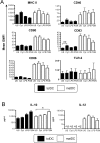
Tolerogenic dendritic cells (tolDCs) have a highly stable semimature, anti-inflammatory phenotype. Phenotypic stability of tolDCs and mature DCs (matDCs) was determined in response to inflammatory stimuli. tolDCs or matDCs were washed and recultured in the absence of tolerising factors including dexamethasone and vitamin D3 for 24 h. Cultures were either unstimulated (US) or stimulated with a cocktail of proinflammatory cytokines (cyt) containing interleukin (IL)-1β, IL-6, tumour necrosis factor (TNF)α (all 10 ng/ml) and interferon (IFN)γ (1000 U/ml), lipopolysaccharide (LPS) (0.1 µg/ml) or peptidoglycan (PGN) (10 µg/ml). (A) DCs were analysed for cell surface phenotype by flow cytometry. Plots show mean geometric mean fluorescence intensity (GMFI) of DC-maturation markers of tolDCs and matDCs. Data are expressed as the mean GMFI±SEM of at least four independent donors. (B) tolDCs and matDCs (105/well) were activated via CD40 by culture with an equivalent number of the J588L-CD40L-expressing cell line in the presence or absence of inflammatory mediators and supernatants collected after 24 h for analysis by ELISA.

References (VSports手机版)

    1. Isaacs JD. T cell immunomodulation – the Holy Grail of therapeutic tolerance. Curr Opin Pharmacol 2007;7:418–25 - "VSports手机版" PubMed
    1. van Duivenvoorde LM, van Mierlo GJ, Boonman ZF, et al. Dendritic cells: vehicles for tolerance induction and prevention of autoimmune diseases. Immunobiology 2006;211:627–32 - PubMed
    1. Banchereau J, Steinman RM. Dendritic cells and the control of immunity. Nature 1998;392:245–52 - PubMed
    1. Ohnmacht C, Pullner A, King SB, et al. Constitutive ablation of dendritic cells breaks self-tolerance of CD4 T cells and results in spontaneous fatal autoimmunity. J Exp Med 2009;206:549–59 - PMC - PubMed
    1. Thomson AW, Robbins PD. Tolerogenic dendritic cells for autoimmune disease and transplantation. Ann Rheum Dis 2008;67(Suppl 3):iii90–6 - PubMed

Publication types

"V体育官网" MeSH terms