<big dropzone="i1UTBJ"><noframes id="U1BBRcjl"> Skip to main page content (VSports在线直播)
U.S. flag

An official website of the United States government

Dot gov

The . gov means it’s official. Federal government websites often end in . gov or . mil. Before sharing sensitive information, make sure you’re on a federal government site. VSports app下载.

Https

The site is secure. The https:// ensures that you are connecting to the official website and that any information you provide is encrypted and transmitted securely. V体育官网.

. 2010 Aug 6;285(32):24529-37.
doi: 10.1074/jbc.M110.127779. Epub 2010 Jun 3.

Characterization of disease-related 5beta-reductase (AKR1D1) mutations reveals their potential to cause bile acid deficiency (V体育官网)

Affiliations

Characterization of disease-related 5beta-reductase (AKR1D1) mutations reveals their potential to cause bile acid deficiency

"V体育官网" Jason E Drury et al. J Biol Chem. .

Abstract

Bile acid deficiency is a serious syndrome in newborns that can result in death if untreated. 5beta-Reductase deficiency is one form of bile acid deficiency and is characterized by dramatically decreased levels of physiologically active 5beta-reduced bile acids VSports手机版. AKR1D1 (aldo-keto reductase 1D1) is the only known human enzyme that stereo-specifically reduces the Delta(4) double bond in 3-keto steroids and sterols to yield the 5beta-hydrogenated product. Analysis of the AKR1D1 gene in five patients with 5beta-reductase deficiency revealed five different mutations resulting in an amino acid substitution in the protein. To investigate a causal role for these observed point mutations in AKR1D1 in 5beta-reductase deficiency, we characterized their effect on enzymatic properties. Attempts to purify mutant enzymes by overexpression in Escherichia coli only yielded sufficient amounts of the P133R mutant for further characterization. This enzyme displayed a highly reduced K(m) and V(max) reminiscent of uncompetitive kinetics with 4-cholesten-7alpha-ol-3-one as substrate. In addition, this mutant displayed no change in cofactor affinity but was more thermolabile in the absence of NADPH as judged by CD spectroscopy. All mutants were compared following expression in HEK 293 cells. Although these enzymes were equally expressed based on mRNA levels, protein expression and functional activity were dramatically reduced. Cycloheximide treatment also revealed that several of the expressed mutants were less stable. Our findings show that the reported mutations in AKR1D1 in patients with 5beta-reductase lead to significantly decreased levels of active enzyme and could be causal in the development of bile acid deficiency syndrome. .


Figures

FIGURE 1.
FIGURE 1.
Location of the disease-related mutants in the AKR1D1 crystal structure. A, position of the mutants on the structure and their relationship to cofactor (magenta) and steroid (blue) binding. B, a perspective view of the location of Pro133 and its relationship to binding testosterone in the unproductive binding mode.
FIGURE 2.
FIGURE 2.
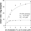
Comparison of velocity versus substrate plots for wild type AKR1D1 and the P133R mutant using Δ4-cholesten-7α-ol-3-one as substrate. ■, AKR1D1; ▴, P133R. Assays were performed using standard fluorimetric assay conditions. For the wild type enzyme, the experiment was replicated twice, and for the P133R mutant, the experiment was replicated once. Measurements were done in duplicate, and variation was less than 10%. One representative experiment is shown.
FIGURE 3.
FIGURE 3.
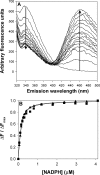
Titration of the fluorescence emission spectra of AKR1D1 with increasing [NADPH]. A, emission spectrum of recombinant AKR1D1 excited at 295 nm following the addition of increasing concentrations of NADPH from 0 to 4 μm. A decrease in emission is seen at 340 nm, and an increase in emission is seen at 460 nm. B, plot of ΔFFmax versus [NADPH], wild type (■), and P133R (▴).
FIGURE 4.
FIGURE 4.
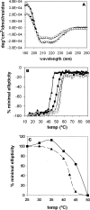
Comparison of the stability of wild type and P133R mutant AKR1D1 using CD spectroscopy and thermal denaturation. A, CD spectra determined for WT AKR1D1 with (□) and without (■) NADPH and AKR1D1-P133R with (Δ) and without (▴) NADPH from 260 to 190 nm. B, melt curve determined for WT AKR1D1 with (□) and without (■) NADPH and AKR1D1-P133R with (Δ) and without (▴) NADPH at 222 nm from 14 to 94 °C. C, activity for WT AKR1D1 (■) and AKR1D1-P133R (▴) heated between 25 and 50 °C for 10 min, followed by a fluorimetric assay with testosterone as substrate. Standard assay conditions were used for measurements. For the heat inactivation experiment shown in C, measurements were performed in duplicate and the mean is given.
FIGURE 5.
FIGURE 5.
Detection of AKR1D1 and AKR1D1 mutant mRNA following transient transfection in HEK 293 cells. HEK 293 cells were transfected with wild type (+Wt) and mutant AKR1D1 (+L106F, +P133R, +P198L, and +R261C), and expression was monitored with primer pairs specifically amplifying either endogenous or vector-expressed (exogenous) AKR1D1. The liver cell line HepG2 expressed AKR1D1 endogenously and served as a positive control for this primer pair. 1D1-pcDNA3.1 is the purified plasmid of the wild type construct transfected into HEK 293 cells and serves as positive control for detection of the exogenous AKR1D1.
FIGURE 6.
FIGURE 6.
Protein expression and stability of AKR1D1 and its natural mutants in transfected HEK 293 cells. Cells were collected at 0, 6, and 24 h following treatment with cycloheximide, and AKR1D1 enzymes were detected by Western blot with a polyclonal anti-AKR1D1 antibody. Blots were stripped and reprobed with antibodies against β-actin to confirm equal loading within an individual experiment. Variations in signal intensity across different constructs are due to differences in exposure time. The arrows point to the signals corresponding to the mutants L106F, P198L, and R261C, that expressed at such low levels that background bands appeared due to increased exposure time.
FIGURE 7.
FIGURE 7.
Detection of 5β-reductase activity of AKR1D1 and its natural mutants in transfected HEK 293 cells. Production of 5β-reduced products from 0.5 μm (A) or 5.0 μm (B) testosterone was followed for up to 48 h in cells expressing either wild type or mutant AKR1D1 or in cells that had been transfected with empty vector (negative control). 5β-Reductase activity for AKR1D1-L106F and AKR1D1-R261C was clearly detectable but close to the detection limit. Cells expressing vector control or AKR1D1-P198L did not exhibit measurable 5β-reduction activity (<0.1 pmol/mg at 60 h of incubation; data not shown). For wild type (C) and AKR1D1-P133R (D), estimates of Km and Vmax were obtained in the transfected cells using testosterone as substrate. All values are normalized to total amount of protein. For A and B, each experiment was performed twice and gave similar results; one representative experiment is shown. For C and D, each point was measured in triplicate with S.E. values less than 10%.

References

    1. Kondo K. H., Kai M. H., Setoguchi Y., Eggertsen G., Sjöblom P., Setoguchi T., Okuda K. I., Björkhem I. (1994) Eur. J. Biochem. 219, 357–363 - PubMed
    1. Deslypere J. P., Young M., Wilson J. D., McPhaul M. J. (1992) Mol. Cell. Endocrinol. 88, 15–22 - PubMed
    1. Wright A. S., Thomas L. N., Douglas R. C., Lazier C. B., Rittmaster R. S. (1996) J. Clin. Invest. 98, 2558–2563 - PMC - PubMed
    1. Bertilsson G., Heidrich J., Svensson K., Asman M., Jendeberg L., Sydow-Bäckman M., Ohlsson R., Postlind H., Blomquist P., Berkenstam A. (1998) Proc. Natl. Acad. Sci. U.S.A. 95, 12208–12213 - PMC - PubMed
    1. Moore L. B., Parks D. J., Jones S. A., Bledsoe R. K., Consler T. G., Stimmel J. B., Goodwin B., Liddle C., Blanchard S. G., Willson T. M., Collins J. L., Kliewer S. A. (2000) J. Biol. Chem. 275, 15122–15127 - PubMed

Publication types

MeSH terms

Substances

LinkOut - more resources