Skip to main page content
U.S. flag

An official website of the United States government

Dot gov

The . gov means it’s official. Federal government websites often end in VSports app下载. gov or . mil. Before sharing sensitive information, make sure you’re on a federal government site. .

Https

The site is secure V体育官网. The https:// ensures that you are connecting to the official website and that any information you provide is encrypted and transmitted securely. .

Comparative Study
. 2010 Jul 1;185(1):263-72.
doi: 10.4049/jimmunol.1000492. Epub 2010 Jun 2.

VSports最新版本 - Early signals during CD8 T cell priming regulate the generation of central memory cells

Affiliations
Comparative Study

V体育平台登录 - Early signals during CD8 T cell priming regulate the generation of central memory cells

Joshua J Obar et al. J Immunol. .

VSports手机版 - Abstract

The CD8(+) T cell response to infection is characterized by the appearance of short-lived (CD127(low) killer cell lectin-like receptor G 1-high) and memory-precursor (CD127(high) killer cell lectin-like receptor G 1-low) effector cells. How and when central-memory T (T(CM); CD62L(high) CCR7(+)) cell and effector-memory T(T(EM); CD62L(low) CCR7(-)) cell subsets are established remains unclear. We now show that the T(CM) cell lineage represents an early developmental branchpoint during the CD8(+) T cell response to infection. Central-memory CD8(+) T cells could be identified prior to the peak of the CD8(+) T cell response and were enriched in lymphoid organs VSports手机版. Moreover, the kinetics and magnitude of T(CM) cell development were dependent on the infectious agent. Furthermore, the extent of early Ag availability, which regulated programmed death-1 and CD25 expression levels, controlled the T(CM)/T(EM) cell lineage decision ultimately through IL-2 and IL-15 signaling levels. These observations identify key early signals that help establish the T(CM)/T(EM) cell dichotomy and provide the means to manipulate memory lineage choices. .


Conflict of interest statement

Disclosures: The authors have no financial conflicts of interest.

Figures

FIGURE 1
FIGURE 1
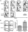
Infection type and turnover rates of TEM cells influence population conversion to TCM cells. A, At the indicated times postinfection of C57BL/6 mice with either VSV-OVA or LM-OVA, the OVA/Kb-specific CD8+ T cells in the spleen, lungs, and lymph nodes were monitored for expression of CD62L. Histograms are gated on OVA/Kb-specific CD8+ T cells. The open histograms show CD62L expression after LM-OVA infection and the filled histograms after VSV-OVA infection. Values in the right corner of each histogram represent the mean percentage of CD62Lhigh cells. Each histogram is representative of four or five mice per time-point and three independent experiments. B, At 30 d after either VSV-OVA or LM-OVA infection, C57BL/6 mice were given BrdU in their drinking water for 4 wk, at which time BrdU incorporation in OVA/Kb-specific CD8+ T cells in the spleen was determined. Filled histograms show BrdU incorporation in CD62Lhigh or CD62Llow OVA/Kb-specific CD8+ T cells, whereas open histograms (dark line) represent an isotype control stain. The graph shows the BrdU incorporation ratio of CD62Lhigh to CD62Llow OVA/Kb-specific memory CD8+ T cells. A value >1.0 indicates that more CD62Lhigh memory cells have incorporated BrdU. Each bar represents the mean value of three mice ± one SD. These data are representative of two independent experiments. Statistical significance was determined using a Student t test. ***p < 0.05.
FIGURE 2
FIGURE 2
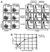
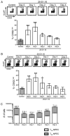
Early postinfection TCM CD8+ T cells are found in the MPEC population. A, C57BL/6 mice were infected i.v. with either VSV-OVA or LM-OVA, and 7 d later the OVA/Kb-specific CD8+ T cell population in the spleen, lungs, and lymph nodes was analyzed. Each effector cell subpopulation, based on KLRG1 and CD127 expression, was assessed for CD62L cell surface expression. The zebra plots are gated on the OVA/Kb-specific CD8+ T cells, whereas the histograms are gated on the respective effector cell subpopulations within the Ag-specific subset. The open histograms show CD62L expression after LM-OVA infection and the filled histograms after VSV-OVA infection. Values in the right corner of each histogram represent the mean percentage of CD62Lhigh cells. These data are representative of four or five mice per group and three independent experiments. B, Expression kinetics of CD62L after i.v. infection with either VSV-OVA or LM-OVA on the CD127high KLRG1low (MPEC) OVA/Kb-specific CD8+ T cell population was monitored in the spleen. The graph represents the proportion of CD127high KLRG1low OVA/Kb-specific CD8+ T cells expressing CD62L. The data presented are the mean of four or five mice per group ± one SD and is representative of two independent experiments.
FIGURE 3
FIGURE 3
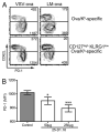
PD-1 expression inversely correlates with CD62L expression of effector cell subsets and is dependent on the strength of TCR engagement. A, C57BL/6 mice were infected i.v. with either VSV-OVA or LM-OVA. At 7 d, expression of PD-1 on both CD62Lhigh and CD62Llow OVA/Kb-specific CD8+ T cells was monitored in the lymph nodes. Contour plots are representative of four or five mice per group and three independent experiments. The top panels are gated on OVA/Kb-specific CD8+ T cells, whereas the bottom panels are further gated on the MPEC pool (CD127high KLRG1low). Values represent the average MFI of PD-1 staining on the CD62Lhigh and CD62Llow populations. Similar data were also obtained from spleen cell analysis. B, C57BL/6 mice were treated i.p. with either 50 μg or 250 μg of the 25-D1.16 Ab or 250 μg of a control Ab (MOPC-21), after which the mice were infected i.v. with VSV-OVA. At 7 d, mice were sacrificed, and expression of PD-1 on the OVA/Kb-specific CD8+ T cells was measured in the spleen. The bar graph is a representation of MFI of PD-1 expression on the CD127high KLRG1low (MPEC) OVA/Kb-specific CD8+ T cells. Data are representative of four or five mice per group and two independent experiments. Statistical significance was determined by a one-way ANOVA analysis. ***p < 0.001; *p < 0.05. MFI, mean fluorescence intensity.
FIGURE 4
FIGURE 4
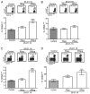
Ag availability during the CD8+ T cell response to infection regulates CD62L expression within the MPEC population. C57BL/6 mice were treated i.p. with either 50 μg or 250 μg of the 25-D1.16 Ab or 250 μg of a control Ab (MOPC-21), after which the mice were infected i.v. with VSV-OVA (A–C) or with LM-OVA (D). At 7 d, the magnitude and phenotype of the OVA/Kb-specific (A, D) or VSV-N/Kb-specific (B) CD8+ T cells were monitored in the spleen. Furthermore, 42 d later, mice were sacrificed, and the magnitude and phenotype of the OVA/Kb-specific CD8+ T cells were monitored in the spleen (C). Dot plots are gated on CD8+ T cells. Values represent the group mean ± one SD. The bar graphs are representations of CD62L expression on the CD127high KLRG1low (MPEC) Ag-specific CD8+ T cells. Data are representative of five mice per group and two independent experiments. Similar data were also observed in the lymph nodes. Statistical significance was determined by a one-way ANOVA analysis. ***p < 0.001; **p < 0.01; *p < 0.05.
FIGURE 5
FIGURE 5
Ag availability early during CD8+ T cell priming regulates CD62L expression within the MPEC population following infection. C57BL/6 mice were treated i.p. with 250 μg of either 25-D1.16 Ab at indicated times or 250 μg of the control Ab (MOPC-21) on day 0. Mice were then infected i.v. with VSV-OVA (A, C) or LM-OVA (B), and 7 d later the OVA/Kb-specific CD8+ T cell population was analyzed in the spleen. Dot plots are gated on CD8+ T cells and display the size of the OVA/Kb-specific CD8+ T cell response. Values represent the group mean ± one SD. The bar graph is a representation of CD62L expression on the CD127high KLRG1low (MPEC) OVA/Kb-specific CD8+ T cells. C shows the ratio of CD62L/CD62L+ cells based on total MPEC numbers. Data are representative of five mice per group and two independent experiments. Similar data were also observed in the lymph nodes. Statistical significance was determined by a one-way ANOVA analysis. ***p < 0.001; **p < 0.01; *p < 0.05.
FIGURE 6
FIGURE 6
Opposing action of IL-2 and IL-15 signaling on regulation of CD62L expression. A, C57BL/6 mice were infected i.v. with 103 CFU of LM-OVA. At the indicated times, expression of CD25 on the OVA/Kb-specific CD8+ T cells was quantified in the spleen by tetramer staining. These data are representative of three to five mice per group and two independent experiments. B, C57BL/6 mice were treated i.p. with 250 μg of either 25-D1.16 or a control mAb (MOPC-21). After this, mice were infected i.v. with 103 CFU of LM-OVA. At 4 d, expression of CD25 on the OVA/Kb-specific CD8+ T cell population in the spleen was analyzed by tetramer enrichment. The filled gray histogram shows CD25 expression on the bulk naive CD8+ T cell population (CD11alow CD44low). The open histograms show CD25 expression on the OVA/Kb-specific CD8+ T cells from control mice (blue line) and 25-D1.16-treated mice (red line). These data are representative of three to five mice and four independent experiments. C, CD25−/− mixed bone marrow chimeras, C57BL/6, and IL-15−/− mice were infected i.v. with 103 CFU of LM-OVA. At 9 d, expression of CD62L on the splenic CD127high KLRG1low (MPEC) OVA/Kb-specific CD8+ T cells was quantified. Data are representative of three to five mice per group and two independent experiments. Statistical significance was measured using a Student t test. *p < 0.05. D, CD25−/− mixed bone marrow chimeras were treated i.p. with 250 μg of either 25-D1.16 or a control mAb (MOPC-21). After this, mice were infected i.v. with 103 CFU of LM-OVA. At 9 d, expression of CD62L on the splenic CD127high KLRG1low (MPEC) OVA/Kb-specific CD8+ T cells was quantified. Data are representative of three to five mice per group and two independent experiments. Statistical significance was measured using a Student t test. **p < 0.01; *p < 0.05.

References

    1. Obar JJ, Khanna KM, Lefrançois L. Endogenous naive CD8+ T cell precursor frequency regulates primary and memory responses to infection. Immunity. 2008;28:859–869. - PMC - PubMed
    1. Kotturi MF, Scott I, Wolfe T, Peters B, Sidney J, Cheroutre H, von Herrath MG, Buchmeier MJ, Grey H, Sette A. Naive precursor frequencies and MHC binding rather than the degree of epitope diversity shape CD8+ T cell immunodominance. J Immunol. 2008;181:2124–2133. - PMC (VSports注册入口) - PubMed
    1. Haluszczak C, Akue AD, Hamilton SE, Johnson LD, Pujanauski L, Teodorovic L, Jameson SC, Kedl RM. The antigen-specific CD8+ T cell repertoire in unimmunized mice includes memory phenotype cells bearing markers of homeostatic expansion. J Exp Med. 2009;206:435–448. - PMC - PubMed
    1. Khanna KM, McNamara JT, Lefrançois L. In situ imaging of the endogenous CD8 T cell response to infection. Science. 2007;318:116–120. - PMC - PubMed
    1. Williams MA, Bevan MJ. Effector and memory CTL differentiation. Annu Rev Immunol. 2007;25:171–192. - PubMed

Publication types

MeSH terms

"V体育2025版" Substances

"V体育官网" LinkOut - more resources