Skip to main page content
U.S. flag

An official website of the United States government

Dot gov

The . gov means it’s official. Federal government websites often end in . gov or VSports app下载. mil. Before sharing sensitive information, make sure you’re on a federal government site. .

Https

The site is secure. The https:// ensures that you are connecting to the official website and that any information you provide is encrypted and transmitted securely. V体育官网.

. 2010 Jun;51(6):1998-2007.
doi: 10.1002/hep.23599.

Accumulation of natural killer T cells in progressive nonalcoholic fatty liver disease

Affiliations

Accumulation of natural killer T cells in progressive nonalcoholic fatty liver disease

Wing-Kin Syn et al. Hepatology. 2010 Jun.

Abstract

Liver inflammation is greater in nonalcoholic steatohepatitis (NASH) than steatosis, suggesting that immune responses contribute to nonalcoholic fatty liver disease (NAFLD) progression. Livers normally contain many natural killer T (NKT) cells that produce factors that modulate inflammatory and fibrogenic responses. Such cells are relatively depleted in steatosis, but their status in more advanced NAFLD is uncertain VSports手机版. We hypothesized that NKT cells accumulate and promote fibrosis progression in NASH. We aimed to determine if livers become enriched with NKT cells during NASH-related fibrosis; identify responsible mechanisms; and assess if NKT cells stimulate fibrogenesis. NKT cells were analyzed in wildtype mice and Patched-deficient (Ptc(+/-)) mice with an overly active Hedgehog (Hh) pathway, before and after feeding methionine choline-deficient (MCD) diets to induce NASH-related fibrosis. Effects of NKT cell-derived factors on hepatic stellate cells (HSC) were examined and fibrogenesis was evaluated in CD1d-deficient mice that lack NKT cells. NKT cells were quantified in human cirrhotic and nondiseased livers. During NASH-related fibrogenesis in wildtype mice, Hh pathway activation occurred, leading to induction of factors that promoted NKT cell recruitment, retention, and viability, plus liver enrichment with NKT cells. Ptc(+/-) mice accumulated more NKT cells and developed worse liver fibrosis; CD1d-deficient mice that lack NKT cells were protected from fibrosis. NKT cell-conditioned medium stimulated HSC to become myofibroblastic. Liver explants were 2-fold enriched with NKT cells in patients with non-NASH cirrhosis, and 4-fold enriched in patients with NASH cirrhosis. .

Conclusion: Hh pathway activation leads to hepatic enrichment with NKT cells that contribute to fibrosis progression in NASH. V体育安卓版.


Figures
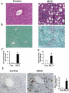
Figure 1
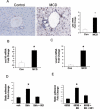
Figure 1. Mice develop NASH-fibrosis when fed a methionine-choline deficient (MCD) diet
Wild-type mice were fed control chow (n=25) or MCD diet (n=25) for 8 weeks, and then sacrificed. (A) Representative hematoxylin & eosin (Final magnification 400X, insert in H&E-stained section from MCD mouse liver shows a ballooned hepatocyte) (B) Sirius red staining after MCD diet (Final magnification 200X).(C) Sirius red quantification by morphometric analysis, and (D) hepatic hydroxyproline content at the end of the treatment period. Results are expressed as fold change relative to control-chow fed mice. (E) αSMA immunoreactivity (final magnification 400X) and (F) αSMA morphometry. Sections from 5 animals were used at each time point and 10 randomly selected, 400X fields chosen for analysis by the Metaview software. Results are expressed as fold change relative to chow-fed control mice and graphed as mean ± SEM. *P<0.05 vs. control mice.
Figure 2
Figure 2. Activation of the Hedgehog-pathway promotes NKT recruitment and static adhesion in murine NASH
Liver tissue from mice described in Figure legend 1 was harvested for QRT-PCR and immunohistochemistry. (A) representative Gli2-staining in MCD diet-treated mice (Final magnification 400X) and mRNA expression of gli2, (B) cxcl16, and (C) vcam-1. Results are expressed as fold change relative to chow-fed control mice, and graphed as mean ± SEM. (D) DN32 static adhesion to immature ductular cells (603B) treated with Sonic hedgehog and / or 5E1, the hedgehog-neutralizing antibody. (E) DN32 static adhesion to 603B cells co-cultured with the myofibroblastic cell line (8B) for 24 hours. Results are expressed as fold change relative to the number of adherent DN32 cells to vehicle-treated ductular cells or ductular cell mono-culture. *P<0.05 vs. control mice. Experiments were performed in duplicate and repeated twice.
Figure 3
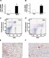
Figure 3. NASH fibrosis is associated with enrichment of liver NKT cells
At the end of 8 weeks, chow-fed control and MCD-fed mice (n=15/group) were sacrificed and QRT-PCR analysis of total liver RNA was done to assess the expression of NKT viability factors. (A) cd1d, and (B) il15 mRNA . Results are expressed as fold change relative to the chow-fed control mice and graphed as mean ± SEM. *P<0.05 vs. control chow-fed mice. (C-D) Additional mice (n=10/group) were treated with control chow or MCD diet for 8 weeks; whole livers were used for FACS analysis. CD4 / CD1d-tetramer double-staining in chow-fed control (C), or MCD-treated mice (D). Livers from all mice were used for FACS analysis. (E-F) CD57 immunoreactive T cells in the hepatic parenchyma of NASH livers (E) compared with normal livers (F) (Final magnification 400X). For each group, sections from 4 mice were analyzed.
Figure 4
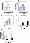
Figure 4. Increased accumulation of intrahepatic NKT cells during diet-induced NASH in Ptc+/- mice with an overly-active Hedgehog-pathway and protection from MCD diet-induced fibrosis in CD1d-deficient mice that lack NKT cells
Ptc+/- mice were fed regular chow (n=2) or MCD diets (n=2) for 8 weeks. At the end of the treatment period, all mice were sacrificed, and intrahepatic mononuclear cells were isolated for FACS analysis. (A) CD4 / CD1d-tetramer double positive cells in chow-fed Ptc+/- mice and (B-C) MCD diet-fed Ptc+/- mice. CD1d-deficient mice and littermate wild type (WT) controls were fed either control chow or MCD diets for 8 weeks (n=3 mice/group/dietary treatment). Fibrosis was evaluated by assessing (E) hepatic hydroxyproline content and performing QRT PCR analysis of (F) hepatic collagen gene expression. Results are expressed as Mean +/- SEM. * P < 0.05, ** P < 0.005
Figure 5
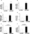
Figure 5. αGalactosylceramide-treated mouse primary liver NKT cells induce primary liver hepatic stellate cell activation
Isolated mouse primary liver mononuclear cells (MNC) (5 × 10⁴) were cultured in complete NKT media, and treated with vehicle or αGalactosylceramide for 24 hours. Cell conditioned-medium were then added to primary cultures of mouse hepatic stellate cells for a further 24 hours; stellate cells were then harvested, and changes in gene expression were assessed by QRT-PCR analysis.(A) gli1, (B) foxf1, (C) collagen 1 α1, (D) αsma, (E) tgfβ, and (F) ctgf. Results are expressed as fold change relative to stellate cells treated with ‘vehicle-MNC’ conditioned-medium. Mean ± SEM of duplicate experiments are graphed. *P<0.05 vs. vehicle-MNC treated stellate cells.
Figure 6
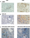
Figure 6. Accumulation of liver NKT cells accompanies Hedgehog-pathway activation in human NASH
(A-C) Liver sections from patients with NASH and either little fibrosis (F0-1) or advanced fibrosis (F3-4) were stained for the marker of activated myofibroblast, αSMA (A), the Hh-regulated NKT chemokine, CXCL16 (B) and NKT cells, i.e., CD3 (blue) / CD57 (brown) double-immunoreactive cells. Representative photomicrographs are shown. Final magnification 400X (A,B) and 600X (C).
Figure 7
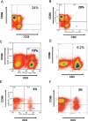
Figure 7. Enrichment of liver mononuclear cells with NKT cells in human cirrhosis
Liver infiltrating leucocytes were isolated from explanted livers of individuals undergoing liver transplantation (n=4), non-diseased liver tissues removed during resection for colorectal hepatic metastases (n=2), as well as excess liver tissues obtained during split-liver grafts (n=2), and analyzed by FACS. (A-B) NASH cirrhosis, (C) hepatitis C cirrhosis, (D) autoimmune hepatitis, and (E-F) non-diseased control livers. NKT cells were identified as CD3 / CD56 double-positive cells.

V体育安卓版 - References

    1. Day CP. Natural history of NAFLD: remarkably benign in the absence of cirrhosis. Gastroenterology. 2005;129:375–378. - PubMed (V体育平台登录)
    1. Adams LA, Lymp JF, St Sauver J, Sanderson SO, Lindor KD, Feldstein A, Angulo P. The natural history of nonalcoholic fatty liver disease: a population-based cohort study. Gastroenterology. 2005;129:113–121. - PubMed
    1. Gao B, Jeong WI, Tian Z. Liver: An organ with predominant innate immunity. Hepatology. 2008;47:729–736. - PubMed
    1. Kronenberg M. Toward an understanding of NKT cell biology: progress and paradoxes. Annu Rev Immunol. 2005;23:877–900. - PubMed
    1. Exley MA, Koziel MJ. To be or not to be NKT: natural killer T cells in the liver. Hepatology. 2004;40:1033–1040. - PubMed

Publication types

MeSH terms

"VSports app下载" LinkOut - more resources