Skip to main page content (VSports)
U.S. flag

An official website of the United States government

Dot gov

The . gov means it’s official. Federal government websites often end in VSports app下载. gov or . mil. Before sharing sensitive information, make sure you’re on a federal government site. .

Https

The site is secure. The https:// ensures that you are connecting to the official website and that any information you provide is encrypted and transmitted securely V体育官网. .

Review
. 2010 Aug;62(8):2206-18.
doi: 10.1002/art.27528.

"VSports" Cartilage cell clusters

Affiliations
Review

Cartilage cell clusters

VSports手机版 - Martin K Lotz et al. Arthritis Rheum. 2010 Aug.

Abstract

The formation of new cell clusters is a histological hallmark of arthritic cartilage but the biology of clusters and their role in disease are poorly understood VSports手机版. This is the first comprehensive review of clinical and experimental conditions associated with cluster formation. Genes and proteins that are expressed in cluster cells, the cellular origin of the clusters, mechanisms that lead to cluster formation and the role of cluster cells in pathogenesis are discussed. .


Figures

Figure 1
Figure 1. Schematic drawing and microscopic image of cell arrangements in normal cartilage
A. Overview of cartilage zones. 1) Each zone has characteristic cell shape, morphology, orientation, and pericellular matrix (PM) deposition. 2) Superficial zone (SZ) cells are small, elongated in shape, parallel to the surface, and lack an extensive PM. These cells predominate the first 50 µm. 3) The middle zone (MZ) is distinguishable by rounded cells that do not exhibit an organized orientation relative to the surface, are within ECM rich in proteoglycans and show presence of PM. 4, 5) Deep zone (DZ) cells show an extensive PM deposition with chondrons in groups of three or more cells arranged in columns perpendicular to the surface. Modified from (138). B. Horizontal sections of normal cartilage. Horizontal sections of normal human articular cartilage were prepared from the SZ to tide mark and stained with safranin O. As indicated on the saggital plane cross-section, image numbers 1–4 were cut at 20µm intervals starting from superficial to middle zone. Image numbers 5–8 represent sections from the MZ, DZ and tidemark at 400µm intervals. Bars: 200µm (large). 50µm (small).
Figure 2
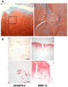
Figure 2. OA clusters
Articular cartilage from a 75-year old male was stained with safranin O (A). The magnification was ×100 (left) and ×400 (right). (B) Articular cartilage from a 57-year old male was stained with ADAMTS-5 and MMP-13. Bar: 200µm (upper) and 50µm (lower).
Figure 3
Figure 3. OA cluster cell apoptosis and calcification
Localization of TUNEL-positive cells, calcium deposits and pyrophosphate-generating enzymes in menisci from OA-affected human knees. The left panel shows apoptotic cells, many in clusters, in the vicinity of (alizarin red-positive) calcified areas. The right panel shows cells immediately bordering calcifications. Staining for plasma cell membrane glycoprotein (PC-1), autotaxin (ATX) and B10 is also prominent at sites of calcification and in areas with TUNEL-positive cells. CILP: cartilage intermediate layer protein. Modified from (131).
Figure 4
Figure 4. OA cluster staining for stem cell markers
A majority of cells in clusters (69 to 79%) are positive for Notch-1, Stro-1 and VCAM-1. Clusters located in the DZ had significantly reduced frequencies of Stro-1 positive cells. (A) Safranin O and Notch-1 staining in clusters (×10 and ×40). (B) Safranin O and Stro-1 staining of OA cartilage sections (×10 and ×40). (C) OA cartilage sections immunostained for VCAM-1 (×10 and ×40). Positive staining indicated by black arrows and negative with white arrows. Modified from (138).
Figure 5
Figure 5. Schematic drawing of mechanisms and phases of cluster formation
(A). Normal cartilage cell organization and resident cartilage progenitors. (B). Injured cartilage with cell death and growth factor release (e.g. FGF2) from disrupted ECM. (C). Further ECM destruction, cell proliferation, cluster formation, cytokine release and abnormal gene expression. Some cells may migrate through DZ or subchondral bone (141).

References

    1. Lawrence RC, Felson DT, Helmick CG, Arnold LM, Choi H, Deyo RA, et al. Estimates of the prevalence of arthritis and other rheumatic conditions in the United States. Part II. Arthritis Rheum. 2008;58(1):26–35. - PMC - PubMed
    1. Loeser RF. Aging and osteoarthritis: the role of chondrocyte senescence and aging changes in the cartilage matrix. Osteoarthritis Cartilage. 2009;17(8):971–979. - PMC - PubMed
    1. Sandell LJ, Aigner T. Articular cartilage and changes in arthritis. An introduction: cell biology of osteoarthritis. Arthritis Res. 2001;3(2):107–113. - PMC - PubMed
    1. Collins . The pathology of articular and spinal diseases. Edward Arnold & Co; 1949. p. 331.
    1. Mankin HJ, Lippiello L. Biochemical and metabolic abnormalities in articular cartilage from osteo-arthritic human hips. J Bone Joint Surg Am. 1970;52(3):424–434. - PubMed

V体育官网 - Publication types

"VSports app下载" MeSH terms