"V体育ios版" Skip to main page content
U.S. flag

An official website of the United States government

Dot gov

The . gov means it’s official. Federal government websites often end in . gov or . mil VSports app下载. Before sharing sensitive information, make sure you’re on a federal government site. .

Https

The site is secure. The https:// ensures that you are connecting to the official website and that any information you provide is encrypted and transmitted securely V体育官网. .

. 2010 May;120(5):1479-93.
doi: 10.1172/JCI41072. Epub 2010 Apr 12.

"VSports最新版本" Altered regulatory T cell homeostasis in patients with CD4+ lymphopenia following allogeneic hematopoietic stem cell transplantation

Affiliations

Altered regulatory T cell homeostasis in patients with CD4+ lymphopenia following allogeneic hematopoietic stem cell transplantation (V体育官网)

Ken-ichi Matsuoka et al. J Clin Invest. 2010 May.

VSports最新版本 - Abstract

CD4+CD25+Foxp3+ Tregs have an indispensable role in the maintenance of tolerance after allogeneic HSC transplantation (HSCT) VSports手机版. Patients with chronic graft-versus-host disease (GVHD) have fewer circulating Tregs, but the mechanisms that lead to this deficiency of Tregs after HSCT are not known. Here, we analyzed reconstitution of Tregs and conventional CD4+ T cells (Tcons) in patients who underwent allogeneic HSCT after myeloablative conditioning. Following transplant, thymic generation of naive Tregs was markedly impaired, and reconstituting Tregs had a predominantly activated/memory phenotype. In response to CD4+ lymphopenia after HSCT, Tregs underwent higher levels of proliferation than Tcons, but Tregs undergoing homeostatic proliferation also showed increased susceptibility to Fas-mediated apoptosis. Prospective monitoring of CD4+ T cell subsets revealed that Tregs rapidly expanded and achieved normal levels by 9 months after HSCT, but Treg levels subsequently declined in patients with prolonged CD4+ lymphopenia. This resulted in a relative deficiency of Tregs, which was associated with a high incidence of extensive chronic GVHD. These studies indicate that CD4+ lymphopenia is a critical factor in Treg homeostasis and that prolonged imbalance of Treg homeostasis after HSCT can result in loss of tolerance and significant clinical disease manifestations. .


Figures

Figure 1
Figure 1. Expression of Foxp3 and suppressive activity of CD4+CD25hiCD127lo Tregs after allogeneic HSCT.
(A) Representative lymphocyte gate for identification of CD4+ T cell subsets is shown. Within the CD4+ T cell gate, Tregs are identified as CD25med-hiCD127lo and Tcons are identified as CD25neg-loCD127med-hi. (B) Gated Tregs and Tcons were examined for intracellular Foxp3 expression. Representative data are shown. Shaded histogram represents isotype control. (C) Tregs or Tcons isolated from patient peripheral blood were cultured with responder Tcons from the same patient and stimulated with irradiated allogeneic PBMCs for 5 days. Method for calculating percentage suppression of proliferation is described in Methods. Data are representative of 5 independent experiments.
Figure 2
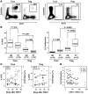
Figure 2. Reduced thymic generation and increased peripheral proliferation of Tregs after allogeneic HSCT.
Gated Tregs and Tcons were examined for CD31 and Ki-67 expression to determine the percentage of RTEs and proliferating cells in each subset. (A) Representative panels for identification of RTEs (left) and proliferating cells (right) are shown. RTEs are identified as CD45RA+CD31+ cells, and proliferating cells are identified as Ki-67+ cells. (B) The percentages of RTEs (left) and proliferating cells (right) in each CD4+ subset from post-HSCT patients (PT) and healthy donors (HD) are shown. Box plots in each figure depict the 75th percentile; median and 25th percentile values and whiskers represent maximum and minimum values. (C) Levels of RTEs (open circles, left y axis) and proliferating cells (closed circles, right y axis) are shown at different times in the first year after HSCT. Results are shown separately for Tcons (left panel) and Tregs (right panel). (D) Correlation of percentage of Ki-67+ proliferating cells in each CD4+ T cell subset and peripheral blood CD4+ T cell count (/μl) of the patient are shown. Tregs (closed triangles) are compared with Tcons (open triangles). Tregs: r = –0.56, P = 0.0008; Tcons: r = –0.32, P = 0.08; Spearman’s test.
Figure 3
Figure 3. Increased expression of Fas (CD95) on CD45RA Tregs is associated with low CD4+ T cell count.
Gated Tregs and Tcons were examined for expression of Fas (CD95). (A) Representative flow cytometry profiles from post-HSCT patients are shown. CD45RA populations in each subset are gated for determination of CD95 expression. (B) Correlation between MFI of CD95 staining on CD45RA Tcons (open triangles) or Tregs (closed triangles) and peripheral blood CD4+ T cell count (cells/μl) for each patient sample are shown. Correlation between the 2 variables was calculated using a rank-based Spearman’s test (Tregs: r = –0.52, P < 0.005; Tcons: r = –0.25, P = 0.19). (C) MFI of CD95 staining is shown for both CD45RA Tcon and Treg subsets from each patient sample. Within each subset, results are compared for samples with high levels of proliferating cells (HP) and low levels of proliferating cells (LP) based on the median percentage of Ki-67 in each sample. The expression of CD95 was significantly greater in high-proliferating cells (%Ki-67+ ≥ 5.0%) than in low-proliferating cells (%Ki-67+ < 5.0%). In contrast, there was no difference in CD95 expression in Tcon samples with either high (%Ki-67+ in ≥ 1.0%) or low levels of proliferation (%Ki-67+ < 1.0%). Results are also compared with values in healthy donors.
Figure 4
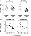
Figure 4. Reduced expression of BCL-2 in CD45RA Tregs undergoing proliferation.
Gated Tregs and Tcons were further subdivided into naive and activated/memory subsets based on the expression of CD45RA. Subsequently, each subset (CD45RA+ Tcons, CD45RA Tcons, CD45RA+ Tregs, and CD45RA Tregs) was examined for intracellular BCL-2 expression. (A) MFI of BCL-2 staining is shown in each subset. Black bars depict median values. (B) Reduced expression of BCL-2 is significantly correlated with percentage of Ki-67 in CD45RA Tregs (right) but not with CD45RA Tcons (left). Correlation between the 2 variables was calculated using a rank-based Spearman’s test (Tregs: r = –0.77, P < 0.01; Tcons: r = –0.18, P = 0.59).
Figure 5
Figure 5. Increased susceptibility to apoptosis and limited proliferative potential in memory Tregs after allogeneic HSCT.
(A) Gating procedure for isolation of CD45RA+ naive Tcons (gated with green), CD45RA memory Tcons (gated with blue), CD45RA+ naive Tregs (gated with orange), and CD45RA memory Tregs (gated with red) by cell sorting. Representative patient sample is shown. (B) Expression of Foxp3 was determined for each CD4+ T cell subset. The results of each subset are shown with respective colors as shown in A. A representative patient sample is shown. Shaded histogram represents isotype control. (C) CFSE-labeled cells were cultured in the presence of anti-CD3, anti-CD28, and IL-2. Cells were harvested after 4 days and stained with anti-CD4 and anti-Foxp3. Gated CD4+ cells were examined. “Alive” cell gate (solid line) and “apoptotic” cell gate (broken line) were defined on the basis of side versus forward scatter (upper panels). Cells in “alive” cell gate were further examined for CFSE dilution and Foxp3 expression (lower panels). A representative result from 5 independent experiments is shown. Numbers denote percentages of cells in the respective quadrants.
Figure 6
Figure 6. Rapid induction of apoptosis in memory Tregs by anti-Fas and inverse correlation with CD4+ T cell count after HSCT.
CD45RA+ Tcons, CD45RA Tcons, CD45RA+ Tregs, and CD45RA Tregs were isolated by cell sorting and cultured separately with anti-Fas antibody or control medium. Apoptosis was assessed by annexin V/7-AAD costaining at several time points, and Fas-specific cell death was calculated as described in Methods. (A) Time-dependent percentage of Fas-specific apoptosis is shown for each CD4+ T cell subset. Data are representative of results from 8 post-HSCT patients and 4 healthy donors. (B) Results of percentage of Fas-specific apoptosis 6 hours after induction are summarized (mean + SEM). *P < 0.01, **P = 0.02. (C and D) Correlation between percentage of Fas-specific apoptosis in each CD4+ subset after 6 hours and peripheral blood CD4+ T cell count (cells/μl) (C) or percentage of CD45RA+ naive population of total CD4+ T cells (D) is shown. Trend lines are shown in respective colors. Correlation between the 2 variables was calculated using a rank-based Spearman’s test. Percentage of Fas-specific apoptosis of memory Tregs was inversely correlated with both CD4+ T cell count and percentage of naive CD4+ T cells (vs. CD4+ T cell counts; r = –0.79, P = 0.02; vs. percentage of naive /CD4+ T cells, r = –0.90, P = 0.002).
Figure 7
Figure 7. Impaired reconstitution of Tregs and increased incidence of chronic GVHD in patients with prolonged severe CD4+ T lymphopenia.
The reconstitution of peripheral blood lymphocyte subsets was assessed prospectively by flow cytometry at 3, 6, 9, 12, 18, and 24 months after HSCT. Median counts (cells/μl) of each subset measured at each time point are shown. (A) Patients were categorized into 2 groups by the absolute number of CD4+ T cells at 6 months after HSCT. 24 patients with CD4+ T cell counts greater than or equal to 260/μl were included in group A and 21 patients with CD4+ T cell counts of less than 260/μl were included in group B. Median CD4+ lymphocyte counts in the each group at each time point are shown. (B) Median Treg counts (solid line, left y axis) and Tcon counts (dashed line, right y axis) in group A and group B patients are shown at serial times over a 2-year period after allogeneic HSCT. Note the scale for Tregs shown on left y axis is one-tenth of the scale for Tcons shown in right y axis. The scales of both left and right y axes for group B are half of the scales shown for group A. (C) Incidence and severity of chronic GVHD are compared for group A (left) and group B (right) patients. (D) Cumulative incidence of extensive chronic GVHD in group A (broken line) and group B (solid line). P value between the 2 groups was calculated using a log-rank test. P = 0.29 at 9 months; P = 0.002 at 24 months.
Figure 8
Figure 8. Altered cytokine milieu during reconstitution of Tregs and Tcons after allogeneic HSCT.
Homeostatic cytokines in plasma were measured at 3, 6, 9, 12, 18, and 24 months after HSCT. (A) Median values (pg/ml) with interquartile range of each cytokine measured at each time point are shown. Gray belt in each graph depicts the interquartile range of healthy controls. IL-7 and IL-15 levels at each time point were significantly higher in patients than in healthy donor controls. *P < 0.003, **P < 0.0001. IL-2 levels in patients were not elevated in the first year. (B) Correlation of plasma cytokine concentration and peripheral blood CD4+ T cell counts (/μl) are shown at 6, 9, and 12 months after transplant. Only IL-15 levels were significantly correlated with CD4+ lymphopenia (Spearman’s test).

V体育安卓版 - References

    1. Bhatia S, et al. Late mortality after allogeneic hematopoietic cell transplantation and functional status of long-term survivors: report from the Bone Marrow Transplant Survivor Study. Blood. 2007;110(10):3784–3792. doi: 10.1182/blood-2007-03-082933. - DOI - PMC - PubMed
    1. Pidala J, Anasetti C, Jim H. Quality of life after allogeneic hematopoietic cell transplantation. Blood. 2009;114(1):7–19. doi: 10.1182/blood-2008-10-182592. - DOI - PMC - PubMed
    1. Ratanatharathorn V, Ayash L, Lazarus HM, Fu J, Uberti JP. Chronic graft-versus-host disease: clinical manifestation and therapy. Bone Marrow Transplant. 2001;28(2):121–129. doi: 10.1038/sj.bmt.1703111. - DOI - PubMed
    1. Lee SJ, Vogelsang G, Flowers ME. Chronic graft-versus-host disease. Biol Blood Marrow Transplant. 2003;9(4):215–233. doi: 10.1053/bbmt.2003.50026. - DOI - PubMed
    1. Sakaguchi S, Sakaguchi N, Asano M, Itoh M, Toda M. Immunologic self-tolerance maintained by activated T cells expressing IL-2 receptor alpha-chains (CD25). Breakdown of a single mechanism of self-tolerance causes various autoimmune diseases. J Immunol. 1995;155(3):1151–1164. - PubMed (VSports最新版本)

Publication types

MeSH terms

"VSports app下载" Substances