<var date-time="vk8tuE"></var> Skip to main page content
U.S. flag

An official website of the United States government

Dot gov

The VSports app下载. gov means it’s official. Federal government websites often end in . gov or . mil. Before sharing sensitive information, make sure you’re on a federal government site. .

Https

The site is secure. The https:// ensures that you are connecting to the official website and that any information you provide is encrypted and transmitted securely. V体育官网.

. 2010 Apr 12;207(4):807-21.
doi: 10.1084/jem.20090348. Epub 2010 Apr 5.

"VSports注册入口" Human cytomegalovirus elicits fetal gammadelta T cell responses in utero

Affiliations

Human cytomegalovirus elicits fetal gammadelta T cell responses in utero

David Vermijlen et al. J Exp Med. .

"VSports最新版本" Abstract

The fetus and infant are highly susceptible to viral infections. Several viruses, including human cytomegalovirus (CMV), cause more severe disease in early life compared with later life VSports手机版. It is generally accepted that this is a result of the immaturity of the immune system. gammadelta T cells are unconventional T cells that can react rapidly upon activation and show major histocompatibility complex-unrestricted activity. We show that upon CMV infection in utero, fetal gammadelta T cells expand and become differentiated. The expansion was restricted to Vgamma9-negative gammadelta T cells, irrespective of their Vdelta chain expression. Differentiated gammadelta T cells expressed high levels of IFN-gamma, transcription factors T-bet and eomes, natural killer receptors, and cytotoxic mediators. CMV infection induced a striking enrichment of a public Vgamma8Vdelta1-TCR, containing the germline-encoded complementary-determining-region-3 (CDR3) delta1-CALGELGDDKLIF/CDR3gamma8-CATWDTTGWFKIF. Public Vgamma8Vdelta1-TCR-expressing cell clones produced IFN-gamma upon coincubation with CMV-infected target cells in a TCR/CD3-dependent manner and showed antiviral activity. Differentiated gammadelta T cells and public Vgamma8Vdelta1-TCR were detected as early as after 21 wk of gestation. Our results indicate that functional fetal gammadelta T cell responses can be generated during development in utero and suggest that this T cell subset could participate in antiviral defense in early life. .


VSports - Figures

Figure 1.
Figure 1.
CMV infection in utero induces an expansion of γδ T cells in newborns. (A) Percentage of γδ T cells of total T cells (CMV+, n = 19; CMV, n = 22). (B) Absolute number of γδ T cells per microliter of cord blood (CMV+, n = 13; CMV, n = 15). (C) Percentage of γδ T cells which are Ki-67+ (CMV+, n = 9; CMV, n = 15) in CMV-infected (gray boxes) and CMV-uninfected (white boxes) newborns. In box-and-whisker graphs, the line at the middle is the median, the box extends from the 25th to 75th percentile, and the error bars, or whiskers, extend down to the lowest value and up to the highest.
Figure 2.
Figure 2.
The expansion of γδ T cells in CMV-infected newborns is restricted to Vγ9 γδ T cells, irrespective of the usage of the Vδ chain. (A) Expression of Vγ9 versus Vδ1, Vδ2, or Vδ3 by γδ T cells from CMV-infected newborn Pos13 (top) and CMV-uninfected newborn Neg4 (bottom). Numbers in dot plots are the percentages of total T cells. (B) Box-and-whisker graph (defined as in Fig. 1) of the percentages of Vγ9+Vδ1+, Vγ9+Vδ2+, Vγ9+Vδ3+, Vγ9Vδ1+, Vγ9Vδ2+, and Vγ9Vδ3+ γδ T cell subpopulations of γδ T cells expressed as a percentage of total T cells. CMV+, n = 10–18; CMV, n = 14–22.
Figure 3.
Figure 3.
Gene expression analysis of γδ T cells derived from three CMV-infected newborns versus γδ T cells derived from three CMV-uninfected newborns. MA plot of differentially expressed genes in γδ T cells upon CMV infection. M (log2 of fold change) reflects the differential expression of a gene. Positive and negative values indicate genes which are up- and down-regulated, respectively, upon CMV infection. A (mean expression) reflects the overall expression level of a gene. Each dot represents one gene. Examples of highly expressed NKR (KIR2DL1), cytotoxic mediators (granzyme B, granzyme H, perforin, and granulysin), cytokines (IFN-γ), and chemokines (CCL4) are indicated.
Figure 4.
Figure 4.
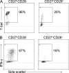
Differentiated (CD27CD28) γδ T cells from CMV-infected newborns express highly the transcription factor T-bet and produce high levels of IFN-γ. Flow cytometry plots for T-bet (A) and IFN-γ (B), gated on CD27CD28 versus gated on CD27+CD28+ γδ T cells of a CMV-positive newborn (Pos12), representative of seven CMV-infected newborns. Cells were stimulated for 4 h with PMA/ionomycin before intracellular staining for IFN-γ. Unstimulated cells showed no IFN-γ staining.
Figure 5.
Figure 5.
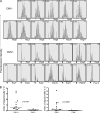
The CDR3δ1 and CDR3δ2 repertoire of γδ T cells from CMV-infected newborns are oligoclonal, and CDR3δ1 is highly enriched for a single sequence. (A) Spectratyping plots of the CDR3δ1 of CMV-uninfected and CMV-infected newborns. Each box represents one donor. The numbers at the left top of each box represents the percentage of Vδ1+ γδ T cells, expressed as percentage of total T cells. The numbers at the right top of each box represent the percentage of CD27CD28 cells of Vδ1+ γδ T cells. The arrows indicate the sequences at CDR3δ1 of 11-aa size of the CMV-infected newborns that have been sequenced: CALGELGDDKLIF (Table S2). (B) Index of oligoclonality for CDR3δ1 and CDR3δ2, determined as described in Materials and methods. Lines indicate medians.
Figure 6.
Figure 6.
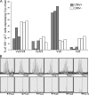
The Vδ1 chain on γδ T cells of CMV-infected newborns preferentially pairs with a public Vγ8 chain. (A) The percentage of Vδ1+ γδ T cells positive for Vγ2/3/4, Vγ5/3, Vγ8, or Vγ9 determined in three CMV-infected (Pos4, Pos6, and Pos13) and three CMV-uninfected newborns. (B) Spectratyping for CDR3γ8 of six CMV-uninfected newborns (top row) and six CMV-infected newborns (bottom row). The arrow indicates the public CDR3γ8 sequence CATWDTTGWFKIF of 11 aa (Table S3). The CDR3γ8 of Pos13 contains 1 aa more (Y).
Figure 7.
Figure 7.
γδ T cell clones expressing the public Vγ8Vδ1 TCR display reactivity against CMV-infected cells via TCR/CD3. Clones were coincubated for 6 h with human embryonic fibroblasts not infected (white bars) or infected (gray bars) with CMV (TB40/E). During coincubation either a control IgG2a antibody (ctrl) or the anti-CD3 antibody OKT3 (anti-CD3) was present in soluble form. Results are shown for one public clone from CMV-infected newborn Pos4 (Vγ8Vδ1-Pos4) and for one public clone of CMV-infected newborn Pos6 (Vγ8Vδ1-Pos6) and are representative of five independent experiments involving 11 different public Vγ8Vδ1 clones.
Figure 8.
Figure 8.
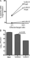
Public Vγ8Vδ1 clones kill CMV-infected target cells and inhibit CMV replication in vitro. (A) CMV-infected (TB40/E) human embryonic fibroblasts were coincubated with either γδ T cell clones expressing the public Vγ8Vδ1 TCR (derived from CMV-infected newborns Pos4 and Pos6) or a control Vγ9Vδ2 clone (derived from a CMV-uninfected newborn) at the indicated effector to target ratios. After 4 h of coincubation, the level of DNA fragmentation in the target cells was quantified. Results are representative of three independent experiments. (B) Human embryonic fibroblasts were incubated with CMV for 2 h, washed, and incubated with medium alone, with a public Vγ8Vδ1 clone from CMV-infected newborn Pos6 or with a control Vγ9Vδ2 clone from a CMV-uninfected newborn. After 7 d, the quantity of infectious CMV from the supernatant was determined by a plaque assay (PFU, plaque forming units). Shown are the mean ± SEM of quadruplicate determinations. Results are representative of two independent experiments.
Figure 9.
Figure 9.
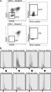
Differentiation and oligoclonal (CALGELGDDKLIF) expansion of fetal γδ T cells can occur early during gestation. (A) Expression of CD27/CD28 and perforin by γδ T cells from a representative CMV-uninfected (bottom) and a representative CMV-infected fetus (Pos4; top), both at the gestational age of 20–21 wk. Dot plots are presented and numbers indicate the percentages of γδ T cells negative for CD27 and CD28 and positive for perforin. CMV, results are representative of 12 (CD27CD28) and 7 (perforin) fetuses (gestation range: 20 wk, 3 d–29 wk, 2 d); CMV+, results are representative of 13 (CD27CD28) and 5 (perforin) fetuses (gestation range: 20 wk, 5 d–29 wk, 2 d). (B) Spectratyping for CDR3δ1 of four CMV-infected fetuses for which we had blood samples both at the time of delivery and at earlier gestation times. The enrichment for the CDR3δ1 size of 11 aa consists of the sequence CALGELGDDKLIF (Table S2).

References

    1. Adams E.J., Chien Y.H., Garcia K.C. 2005. Structure of a gammadelta T cell receptor in complex with the nonclassical MHC T22. Science. 308:227–231 10.1126/science.1106885 - VSports手机版 - DOI - PubMed
    1. Andrade F., Fellows E., Jenne D.E., Rosen A., Young C.S. 2007. Granzyme H destroys the function of critical adenoviral proteins required for viral DNA replication and granzyme B inhibition. EMBO J. 26:2148–2157 10.1038/sj.emboj.7601650 - DOI - PMC - PubMed
    1. Appay V., Dunbar P.R., Callan M., Klenerman P., Gillespie G.M., Papagno L., Ogg G.S., King A., Lechner F., Spina C.A., et al. 2002. Memory CD8+ T cells vary in differentiation phenotype in different persistent virus infections. Nat. Med. 8:379–385 10.1038/nm0402-379 - DOI - PubMed
    1. Battistini L., Borsellino G., Sawicki G., Poccia F., Salvetti M., Ristori G., Brosnan C.F. 1997. Phenotypic and cytokine analysis of human peripheral blood gamma delta T cells expressing NK cell receptors. J. Immunol. 159:3723–3730 - PubMed
    1. Beldjord K., Beldjord C., Macintyre E., Even P., Sigaux F. 1993. Peripheral selection of Vδ1+ cells with restricted T cell receptor δ gene junctional repertoire in the peripheral blood of healthy donors. J. Exp. Med. 178:121–127 10.1084/jem.178.1.121 - DOI - PMC - PubMed

"V体育官网入口" Publication types

MeSH terms