Skip to main page content
U.S. flag

An official website of the United States government

Dot gov

The . gov means it’s official. Federal government websites often end in . gov or . mil. Before sharing sensitive information, make sure you’re on a federal government site VSports app下载. .

Https

The site is secure V体育官网. The https:// ensures that you are connecting to the official website and that any information you provide is encrypted and transmitted securely. .

. 2010 Apr 5;189(1):111-26.
doi: 10.1083/jcb.200902153. Epub 2010 Mar 29.

Caveolin-1-dependent occludin endocytosis is required for TNF-induced tight junction regulation in vivo

Affiliations

Caveolin-1-dependent occludin endocytosis is required for TNF-induced tight junction regulation in vivo

"VSports最新版本" Amanda M Marchiando et al. J Cell Biol. .

Abstract

Epithelial paracellular barrier function, determined primarily by tight junction permeability, is frequently disrupted in disease. In the intestine, barrier loss can be mediated by tumor necrosis factor (alpha) (TNF) signaling and epithelial myosin light chain kinase (MLCK) activation. However, TNF induces only limited alteration of tight junction morphology, and the events that couple structural reorganization to barrier regulation have not been defined. We have used in vivo imaging and transgenic mice expressing fluorescent-tagged occludin and ZO-1 fusion proteins to link occludin endocytosis to TNF-induced tight junction regulation VSports手机版. This endocytosis requires caveolin-1 and is essential for structural and functional tight junction regulation. These data demonstrate that MLCK activation triggers caveolin-1-dependent endocytosis of occludin to effect structural and functional tight junction regulation. .


Figures

Figure 1.
Figure 1.
Occludin endocytosis begins 90 min after TNF administration and precedes intestinal fluid accumulation. (A) Jejunum was harvested from wild-type mice at the indicated times after intraperitoneal injection of 5 µg TNF and labeled for claudin proteins or E-cadherin (green), ZO-1 or F-actin (red), and nuclei (blue). (B) As in A, jejunal sections were labeled for occludin (green), F-actin (red), and nuclei (blue). (C) Jejunal sections were harvested and labeled as in B. TNF treatment leaves large regions of the tight junction completely lacking in occludin (arrows). (D) Jejunal epithelia were isolated from wild-type mice 120 min after injection with saline or TNF and analyzed via immunoblotting. (E) Number of occludin-containing vesicles (black circles) was assessed morphometrically, and fluid accumulation (white boxes) was measured as weight/length ratio (n = 4). Error bars indicate mean ± SEM. Bars, 10 µm.
Figure 2.
Figure 2.
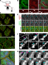
In vivo imaging of EGFP-occludin endocytosis in jejunal enterocytes. (A) The opened jejunum, with intact neurovascular supply, is placed in a dish with the mucosa resting on a coverslip. (B) EGFP-occludin (green) is targeted to tight junctions and, to a lesser degree, lateral membranes of jejunal enterocytes. Villous capillaries within the lamina propria can be identified by confocal reflectance (red) based on the presence of blood flow. Portions of four villi are shown in cross section. Nuclei (blue) are shown in the merged image. (bottom) High speed imaging shows erythrocytes (arrows) flowing through villus capillaries (outlined by dashed lines). Images are taken from Video 1. Bars: (top) 20 µm; (bottom) 5 µm. (C) XY plane images at the indicated relative z positions show the targeting of mRFP1–ZO-1 (red) and EGFP-occludin (green) in jejunal enterocytes. Both proteins are concentrated at the tight junction. EGFP-occludin is also present in lateral membranes. The full z stack is shown in Video 2. Bar, 20 µm. (D) 125 confocal sections collected at 0.1-µm intervals were used to create this reconstruction of three villi from a transgenic mouse expressing mRFP1–ZO-1 (red) and EGFP-occludin (green). Nuclei are blue. A rotating view of the reconstruction is available as Video 3. Bar, 20 µm. (E) High magnification images of villous epithelium show that mRFP1–ZO-1 (top; red in merge) and EGFP-occludin (middle; green in merge) were collected at the plane of the tight junction as determined by the location of mRFP1–ZO-1 (pink arrows). EGFP-occludin endocytosis occurs during the interval from 85 to 185 min after TNF injection (blue arrows). The complete time-lapse series is shown in Video 4. Bar, 10 µm. (F) The relatively low magnification image is shown for orientation. Images were collected from a small area of the tight junction (pink arrows). The higher magnification images of the boxed area show focal EGFP-occludin enrichment before endocytosis, vesicle budding, separation, and movement out of the focal plane (blue arrows). The entire process takes ∼15 min. Time after TNF injection is indicated. Bars: (left) 5 µm; (middle) 1 µm. (G) The lower magnification image is shown for orientation. The tight junction (pink arrows) appears as a bright spot of EGFP-occludin. Higher magnification images of the boxed area show EGFP-occludin–containing endocytic vesicles (blue arrows) leaving the basal aspect of the tight junction. Time after TNF injection is indicated. Bars: (left) 10 µm; (middle) 2 µm.
Figure 3.
Figure 3.
EGFP-occludin mice are partially protected from TNF-induced occludin redistribution, barrier dysfunction, and fluid secretion. (A) Jejunal epithelia were isolated from wild-type and EGFP-occludin transgenic mice. Occludin, EGFP-occludin, and actin content were assessed by immunoblotting. (B) Jejunum was harvested from wild-type and EGFP-occludin mice 120 min after TNF injection. Wild-type tissue was labeled for occludin (top; green in merge) and F-actin (red). EGFP-occludin (top; green in merge) and labeled F-actin (red) are shown for transgenic animals. Regions of the tight junction completely lacking in occludin (arrows) develop in TNF-treated wild-type but not EGFP-occludin transgenic mice. Bar, 10 µm. (C) In vivo perfusion assays were used to assess paracellular BSA flux in wild-type mice and EGFP-occludin mice injected with TNF (gray bars) or vehicle (white bars; n = 6). (D) In vivo perfusion assays of water movement in wild-type and EGFP-occludin mice injected with TNF (gray bars) or vehicle (white bars; n = 6). Error bars indicate mean ± SEM.
Figure 4.
Figure 4.
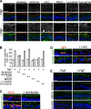
Inhibitors of caveolar endocytosis prevent TNF-induced occludin internalization. (A) Wild-type mice were injected with vehicle or TNF as indicated. A segment of jejunum was perfused with 50 µM dynasore, 60 µM amiloride, 200 µM chlorpromazine (CPZ), 2 mM MβCD, 50 µM L-t-LacCer, or 50 µM D-e-LacCer and harvested 135 min later. Sections were labeled for occludin (grayscale images; green in merge), F-actin (red), and nuclei (blue). Bar, 20 µm. (B) Morphometric analysis of the number of occludin-containing vesicles per enterocyte in wild-type mice injected with vehicle (white bars) or TNF (gray bars) in jejunal segments perfused with saline or the indicated inhibitors. (C) Wild-type mice were injected with TNF, and a segment of jejunum perfused with Alexa Fluor 594–conjugated WGA (50 µg/ml). Perfused segments were harvested 135 min after TNF treatment. Sections were labeled for actin (top; green) or occludin (bottom; green) and nuclei (blue). Perfusion with amiloride prevented endocytosis of WGA (red) but not occludin. Bar, 10 µm. (D) Wild-type mice were injected with TNF, and a segment of jejunum perfused with DyLight 594–conjugated IgG (40 µg/ml). Perfused segments were harvested 135 min after TNF treatment. Sections were labeled for actin (top; green) or occludin (bottom; green) and nuclei (blue). Perfusion with chlorpromazine prevented endocytosis of IgG (red) but not occludin. Bar, 10 µm. (E) Sections of jejunum from mice injected with vehicle or TNF and perfused with saline, dynasore, MβCD, or L-t-LacCer harvested 135 min after TNF injection were labeled for phosphorylated MLC (green) and nuclei (blue). Bar, 20 µm. Error bars indicate mean ± SEM.
Figure 5.
Figure 5.
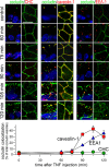
TNF induces colocalization of occludin and caveolin-1 but not clathrin heavy chain. Wild-type mice were injected with vehicle or TNF, and jejunum harvested at the times indicated. Sections were labeled for occludin (green), nuclei (blue), and clathrin heavy chain, caveolin-1, or EEA1 (all red). Apical–basal-oriented and orthogonal sections are shown. Morphometric analysis of the fraction of occludin-containing vesicles that also contain caveolin-1 (red squares), clathrin heavy chain (green circles), and EEA1 (blue triangles) is shown below the micrographs. Error bars indicate mean ± SEM. Bars, 10 µm.
Figure 6.
Figure 6.
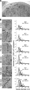
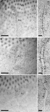
Distinct vesicle populations are impacted by TNF. Electron micrographs of aldehyde-fixed, plastic-embedded jejunum from control and TNF-treated wild-type mice are shown. (A) Low magnification view of jejunal enterocytes showing the relationship between the intestinal lumen (L), microvillus brush border (Mv), tight junction (TJ), adherens junction (AJ), and desmosomes (D). Note the exclusion of mitochondria (M) from the most apical cytoplasm by the dense perijunctional actomyosin ring (PAMR). Bar, 2 µm. (B) Jejunal enterocytes of untreated mice (0 min) and mice sacrificed at the indicated times after TNF treatment were examined. Representative electron micrographs are shown. At least 1,000 µm2 apical cytoplasm was examined per condition. Incremental fits show that the data can be modeled as the sum of four Gaussian distributions (solid lines). Mean diameters of 80, 125, 170, and 240 nm (dashed lines) are shown. The number of vesicles in each population (per 1,000 µm2 cytoplasm) is indicated above each curve. The actual number of vesicles observed (per 1,000 µm2 cytoplasm) is shown in the top right corner of each graph. Bar, 500 nm.
Figure 7.
Figure 7.
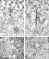
Immunogold labeling of occludin and caveolin-1 in jejunal enterocytes of untreated wild-type mice. High pressure–frozen, freeze-substituted, cryoembedded specimens were immunolabeled. (A) Antioccludin, detected with 10 nm gold-conjugated secondary antisera, shows tight junction–specific labeling (best appreciated in the enlarged region [right]). (B) Anti–caveolin-1, detected with 10 nm gold-conjugated secondary antisera, shows labeling at the adherens junction (AJ) and, to a lesser extent, the tight junction (TJ). (C) Antioccludin and anti–caveolin-1, detected with 10 nm and 15 nm gold-conjugated secondary antisera, respectively, label at the tight junction and adherens junction as in A and B. Controls for double labeling are shown in Fig. S1. Mv, microvilli. Bars: (left) 300 nm; (right) 50 nm.
Figure 8.
Figure 8.
Immunogold labeling of occludin and caveolin-1 in jejunal enterocytes of 90-min TNF-treated wild-type mice. High pressure–frozen, freeze-substituted, cryoembedded specimens were immunolabeled with antioccludin and anti–caveolin-1, which were detected with 10 nm and 15 nm gold-conjugated secondary antisera, respectively. (A and B) Occludin and caveolin-1 colocalize at tight junctions (TJ) and adherens junctions (AJ). Mv, microvilli. (C and D) The population of vesicles (V) formed after TNF treatment contains occludin and caveolin-1. Single-label images are shown in Fig. S2. Bars, 300 nm.
Figure 9.
Figure 9.
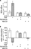
Inhibition of caveolar endocytosis prevents TNF-induced barrier dysfunction and fluid secretion. (A) In vivo perfusion assays of wild-type mice injected with TNF (gray bars) or vehicle (white bars). Inclusion of 50 µM dynasore or 2 mM MβCD within the perfusion solution prevented TNF-induced increases in BSA flux (n = 3). (B) The direction of water movement is reversed from net absorption to net secretion in wild-type mice injected with TNF (gray bars) relative to wild-type mice treated with vehicle (white bars). Dynasore prevented and MβCD reduced TNF-induced water secretion (n = 3). Error bars indicate mean ± SEM.
Figure 10.
Figure 10.
Caveolin-1 is required for TNF-induced occludin internalization, barrier dysfunction, and net water secretion. (A) Caveolin-1+/+ and caveolin-1−/− mice were injected with vehicle or TNF as indicated, and a segment of jejunum harvested 135 min later. Sections were labeled for occludin (green), F-actin (red), and nuclei (blue). Bar, 10 µm. (B) Electron micrographs demonstrate TNF-induced perijunctional actomyosin condensation in jejunal enterocytes of caveolin-1+/+ and caveolin-1−/− mice. Bar, 125 nm. (C) Jejunal epithelial cells were isolated from caveolin-1+/+ and caveolin-1−/− mice 135 min after injection with vehicle or TNF. Cell lysates were analyzed by immunoblotting for phosphorylated and total MLC. (D) Jejunum was harvested 135 min after caveolin-1+/+ and caveolin-1−/− mice were injected with vehicle or TNF. Sections were labeled for phosphorylated MLC (green) and nuclei (blue). Bar, 10 µm. (E) Mucosal TNF mRNA transcripts measured by quantitative RT-PCR were increased by TNF injection in jejunum of caveolin-1+/+ and caveolin-1−/− mice. (F) Cell lysates were analyzed by immunoblotting for occludin, caveolin-1, clathrin heavy chain, and E-cadherin. Protein content was not affected by acute TNF exposure. (G) In vivo perfusion assays show that TNF increases paracellular flux in caveolin-1+/+ mice but not in caveolin-1−/− mice. (H) TNF reverses the direction of water movement from net absorption to net water secretion in caveolin-1+/+ but not caveolin-1−/− mice (n = 6). Error bars indicate mean ± SEM.

References

    1. Akiba Y., Furukawa O., Guth P.H., Engel E., Nastaskin I., Sassani P., Dukkipatis R., Pushkin A., Kurtz I., Kaunitz J.D. 2001. Cellular bicarbonate protects rat duodenal mucosa from acid-induced injury. J. Clin. Invest. 108:1807–1816 - PMC - PubMed
    1. Balda M.S., Whitney J.A., Flores C., González S., Cereijido M., Matter K. 1996. Functional dissociation of paracellular permeability and transepithelial electrical resistance and disruption of the apical-basolateral intramembrane diffusion barrier by expression of a mutant tight junction membrane protein. J. Cell Biol. 134:1031–1049 10.1083/jcb.134.4.1031 - DOI - PMC - PubMed
    1. Bentzel C.J., Hainau B., Edelman A., Anagnostopoulos T., Benedetti E.L. 1976. Effect of plant cytokinins on microfilaments and tight junction permeability. Nature. 264:666–668 10.1038/264666a0 - DOI - PubMed
    1. Berglund J.J., Riegler M., Zolotarevsky Y., Wenzl E., Turner J.R. 2001. Regulation of human jejunal transmucosal resistance and MLC phosphorylation by Na(+)-glucose cotransport. Am. J. Physiol. Gastrointest. Liver Physiol. 281:G1487–G1493 - PubMed (V体育2025版)
    1. Blair S.A., Kane S.V., Clayburgh D.R., Turner J.R. 2006. Epithelial myosin light chain kinase expression and activity are upregulated in inflammatory bowel disease. Lab. Invest. 86:191–201 10.1038/labinvest.3700373 - DOI - PubMed

Publication types

MeSH terms