Skip to main page content (VSports)
U.S. flag

An official website of the United States government

Dot gov

The . gov means it’s official. Federal government websites often end in . gov or . mil. Before sharing sensitive information, make sure you’re on a federal government site. VSports app下载.

Https

The site is secure. The https:// ensures that you are connecting to the official website and that any information you provide is encrypted and transmitted securely. V体育官网.

. 2010 Mar 15;184(6):3284-97.
doi: 10.4049/jimmunol.0902199. Epub 2010 Feb 17.

A distinct subset of proinflammatory neutrophils isolated from patients with systemic lupus erythematosus induces vascular damage and synthesizes type I IFNs

Affiliations

A distinct subset of proinflammatory neutrophils isolated from patients with systemic lupus erythematosus induces vascular damage and synthesizes type I IFNs

Michael F Denny et al. J Immunol. .

Erratum in

  • J Immunol. 2010 Sep 15;185(6):3779

Abstract

Neutrophil-specific genes are abundant in PBMC microarrays from lupus patients because of the presence of low-density granulocytes (LDGs) in mononuclear cell fractions. The functionality and pathogenicity of these LDGs have not been characterized. We developed a technique to purify LDGs from lupus PBMCs and assessed their phenotype, function, and potential role in disease pathogenesis. LDGs, their autologous lupus neutrophils, and healthy control neutrophils were compared with regard to their microbicidal and phagocytic capacities, generation of reactive oxygen species, activation status, inflammatory cytokine profile, and type I IFN expression and signatures. The capacity of LDGs to kill endothelial cells and their antiangiogenic potential were also assessed. LDGs display an activated phenotype, secrete increased levels of type I IFNs, TNF-alpha, and IFN-gamma, but show impaired phagocytic potential. LDGs induce significant endothelial cell cytotoxicity and synthesize sufficient levels of type I IFNs to disrupt the capacity of endothelial progenitor cells to differentiate into mature endothelial cells. LDG depletion restores the functional capacity of endothelial progenitor cells. We conclude that lupus LDGs are proinflammatory and display pathogenic features, including the capacity to synthesize type I IFNs. They may play an important dual role in premature cardiovascular disease development in systemic lupus erythematosus by simultaneously mediating enhanced vascular damage and inhibiting vascular repair. VSports手机版.


Conflict of interest statement

The authors have no conflicting financial interests.

Figures
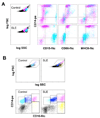
Figure 1
Figure 1. Identification of LDGs in lupus PBMC fractions
Healthy control or SLE PBMCs were stained for markers of the monocyte or granulocyte lineages and analyzed by FACS. A. Gates which contained predominantly lymphocytes, monocytes and granulocytes were established in dual-log scattergrams. Granulocytes (blue) and monocytes (pink) are distinguished based on CD14, CD15, CD86 and MHC class II expression. Monocytes express high levels of CD14 and are positive for CD86 and MHC class II, while CD15 is weak or absent. Granulocytes present in the PBMC fraction are CD15hi, CD14lo and negative for CD86 and MHC class II. Similar results were seen in 2 additional controls and 5 additional SLE patients. B. Analysis of CD86 and CD16 revealed several subpopulations. Most healthy control monocytes display the resting phenotype of CD86+CD16 (light blue), while SLE monocytes have the more activated phenotype of CD86+CD16+ (blue). The CD16hi cells can be further divided based on CD86 expression. The CD16hi/CD86 pool (yellow) likely represents LDGs, while the CDhi/CD86+ population (pink) possibly reflects conjugates of CD16hi granulocytes and CD86+ monocytes.
Figure 2
Figure 2. Enrichment of lupus LDGs
LDGs were isolated from lupus PBMC fractions by negative selection with magnetic beads. A. With this approach highly enriched granulocytes were obtained. The cells express granulocyte markers (high levels of CD15, CD16, CD11c, CD14 and CD10). The position of the isotype control Ab is shown in the gray histogram. Similar results were seen in 2 additional SLE patients. B. Enriched granulocytes were stained with CD16-FITC and CD15-biotin followed by Cy3-streptavidin; nuclei were counterstained with Hoechst 33342. Surface expression and co-distribution of CD15 and CD16 is apparent (original total magnification 1000x). Similar result was seen in one additional SLE patient. C. Differential staining of the enriched granulocyte fraction obtained from lupus PBMCs reveals that the cells are neutrophils and have a range of nuclear morphologies. Segmented (Seg) and band neutrophils are apparent, as well as myelocyte-like cells (total magnification 1000×). Similar results were seen in 3 additional SLE patients.
Figure 3
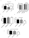
Figure 3. Neutrophil function of LDGs
A. Phagocytic capacity of bacteria is impaired in lupus LDGs. Results represent mean± SEM fluorescence of 5 control and 5 lupus patients each performed in triplicate, as assessment of phagocytosis of S. aureus particles. P values are included. B. Bactericidal activity is preserved in LDGs. Bar graph displays the percentage S.aureus CFU decrease observed between 10 to 40 min and 10 to 70 min after the addition of lysostaphin in lupus LDGs, autologous lupus neutrophils or control neutrophils. Results represent the mean ± SEM from 5 lupus and 5 control patients, each performed in triplicate; p=not significant between groups. C. Intracellular MPO levels in LDGs are similar to normal density neutrophils. Results represent the mean±SEM mean fluorescent intensity of intracellular MPO in LDGs and neutrophils (5 control and 5 lupus patients; p=not significant). D. LDGs do not differ from neutrophils in hydrogen peroxide synthesis capacity after PMA stimulation. Results represent mean±SEM H2O2 concentration (µM/105 cells/ 60 min) of 5 control and 5 lupus samples, each performed in triplicate; p=not significant. E. LDGs do not differ from neutrophils in hydrogen peroxide synthesis capacity after immune complex stimulation. Results represent mean±SEM H2O2 concentration (µM/105 cells/ 45 min) of 5 control and 5 lupus samples, each performed in triplicate; p=not significant.
Figure 4
Figure 4. LDGs secrete increased levels of proinflammatory cytokines
Results represent A- eicosanoid and B–D- cytokine concentration in cell supernatants in pg/mL. Results are shown using unstimulated or PMA-stimulated cells and measurements were done with supernatants harvested after 48 h in culture.; * P<0.05 when comparing LDGs to autologous neutrophils and control neutrophils and ** P<0.05 when comparing LDGs to control neutrophils. Results represent mean±SEM of 10 controls and 10 SLE patients.
Figure 5
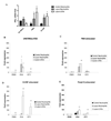
Figure 5. Lupus LDGs express increased levels of IFN-α mRNA and their supernatants induce enhanced levels of type I IFN-inducible genes
A. Results represent mean±SEM of IFN-α mRNA expression, after adjusting for housekeeping gene (HPRT1). Results are from 4 controls and 5 SLE patients. B–D. Results represent fold induction of IFN-inducible genes (mRNA) on epithelial cell lines by neutrophil supernatants and are presented as mean ± SEM fold-induction by supernatants from control neutrophils (n=5), SLE neutrophils or autologous lupus LDGs (n=8 each). Data are normalized to HPRT-1, *p<0.05 when comparing to control neutrophils and **p<0.05 when comparing LDGs to both control and lupus neutrophils.
Figure 6
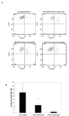
Figure 6. Lupus LDGs are cytotoxic to the endothelium
A. Results show representative dot plots of endothelial cell apoptosis induced by LDGs, autologous lupus neutrophils or control neutrophils. Neutrophils representing CD10+ cells are excluded from analysis. CD146+ cells represent endothelial cells and Annexin V + cells are considered apoptotic or early necrotic. Numbers represent % expression in the various quadrants. Apoptotic endothelial cells are CD146+Annexin V+. B. Bar graphs represent % of apoptotic HUVECs after overnight exposure to LDGs, autologous lupus neutrophils or control neutrophils. Results represent mean +SEM of 7 SLE and 9 controls samples, *p<0.05 when comparing lupus neutrophils to control neutrophils and ** p<0.01 when comparing LDGs to autologous lupus neutrophils and to control neutrophils.
Figure 7
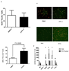
Figure 7. LDG depletion improves the capacity of EPCs/CACs to become mature endothelial cells by abrogating type I IFN activity
Lupus PBMCs were cultured under proangiogenic conditions and incubated at different time-points during culture with diI–acetylated LDL and FITC–UEA-1. Mature endothelial cells were identified by co-expression of these markers. A. Bar graphs represent number of mature endothelial cells per power field at day 7, when comparing initial plating of unfractionated lupus PBMCs versus PBMCs depleted of pDCs (pDC(−)) (top) or LDGs (CD10 depletion, CD10(−)) (bottom). Results are mean± SEM of 4 independent experiments for pDC depletion and 6 independent experiments for LDG depletion; p=NS for pDC depletions and p=0.0049 for LDG depletion. B. Results are representative images of mature endothelial cells obtained after 7 days in culture, when comparing initial plating of lupus unfractionated PBMCs versus PBMCs depleted of either pDCs (top) or LDGs (bottom) exposed to proangiogenic stimulation. Lupus CD10 depletion, but not pDC depletion, resulted in enhanced numbers of mature endothelial cells. C. Bar graphs represent the capacity of supernatants obtained at day 7 from lupus unfractionated PBMCs, PBMCs depleted of either pDCs or LDGs (all cultured under proangiogenic stimulation) to induce type I IFN-inducible genes in epithelial cell lines. Results are mean± SEM of 4 independent experiments for pDC depletion and 6 independent experiments for LDG depletion; *p<0.05 when comparing type I IFN induction between total PBMCs and CD10-depleted PBMC cultures.

References

    1. Kyttaris VC, Juang YT, Tsokos GC. Immune cells and cytokines in systemic lupus erythematosus: an update. Curr Opin Rheumatol. 2005;17:518–522. - PubMed
    1. Bennett L, Palucka AK, Arce E, Cantrell V, Borvak J, Banchereau J, Pascual V. Interferon and granulopoiesis signatures in systemic lupus erythematosus blood. J Exp Med. 2003;197:711–723. - PMC - PubMed
    1. Denny MF, Chandaroy P, Killen PD, Caricchio R, Lewis EE, Richardson BC, Lee KD, Gavalchin J, Kaplan MJ. Accelerated macrophage apoptosis induces autoantibody formation and organ damage in systemic lupus erythematosus. J Immunol. 2006;176:2095–2104. - PubMed
    1. Banchereau J, Pascual V. Type I interferon in systemic lupus erythematosus and other autoimmune diseases. Immunity. 2006;25:383–392. - PubMed
    1. Kaplan MJ, Lewis EE, Shelden EA, Somers E, Pavlic R, McCune WJ, Richardson BC. The apoptotic ligands TRAIL, TWEAK, and Fas ligand mediate monocyte death induced by autologous lupus T cells. J Immunol. 2002;169:6020–6029. - PubMed

"V体育2025版" Publication types

MeSH terms

"V体育平台登录" Substances