Skip to main page content
U.S. flag

An official website of the United States government

Dot gov

The . gov means it’s official. Federal government websites often end in . gov or . mil. Before sharing sensitive information, make sure you’re on a federal government site VSports app下载. .

Https

The site is secure. The https:// ensures that you are connecting to the official website and that any information you provide is encrypted and transmitted securely. V体育官网.

. 2010 Aug;12(8):776-89.
doi: 10.1093/neuonc/noq003. Epub 2010 Feb 17.

VSports手机版 - Analysis of receptor tyrosine kinases (RTKs) and downstream pathways in chordomas

Affiliations

V体育2025版 - Analysis of receptor tyrosine kinases (RTKs) and downstream pathways in chordomas

Elena Tamborini et al. Neuro Oncol. 2010 Aug.

"V体育平台登录" Abstract

We have previously demonstrated that chordomas express activated platelet-derived growth factor receptor (PDGFRB) and that treatment with imatinib, which is capable of switching off the activation of various receptor tyrosine kinases (RTKs) including PDGFRB, benefits a number of patients. The aim of this study was to identify the possible presence of other activated RTKs and their downstream signaling effectors. Cryopreserved material from 22 naïve sporadic chordomas was investigated for the presence of activated RTKs and their cognate ligands and downstream signaling effectors by means of human phospho-RTK antibody arrays, Western blotting, and molecular analysis; immunohistochemistry and fluorescence in situ hybridization were used to analyze the corresponding formalin-fixed and paraffin-embedded samples. We detected activated PDGFRB, FLT3, and colony stimulating factor 1 receptor (CSF1R) of the PDGFR family and highly phosphorylated EGFR, HER2/neu, and (to a lesser extent) HER4 of the EGFR family VSports手机版. The detection of PDGFRB/PDGFB confirmed our previous data. The presence of activated EGFR was paralleled by the finding of high levels of epidermal growth factor (EGF) and transforming growth factor alpha (TGFalpha) and PDGFB co-expression and PDGFRB co-immunoprecipitation. Of the downstream effectors, the PI3K/AKT and RAS/MAPK pathways were both activated, thus leading to the phosphorylation of mammalian target of rapamycin (mTOR) and 4E-BP1 among the regulators involved in translational control. Taken together, our results (i) provide a rationale for tailored treatments targeting upstream activated receptors, including the PDGFR and EGFR families; (ii) support the idea that a combination of upstream antagonists and mTOR inhibitors enhances the control of tumor growth; and (iii) indicate that the 4E-BP1/eIF4E pathway is a major regulator of protein synthesis in chordoma. .


Figures

Fig. 1.
Fig. 1.
Usual regulation of RTK downstream effectors. The grey lines indicate new pathways related to protein activation/inhibition by means of phosphorylation (thick continuous lines) or transcriptional silencing (dotted lines). In addition to being phosphorylated by p70S6K/mTOR, S6 may be activated by p90RSK. The same is true of 4E-BP1 which, in addition to mTOR, may be phosphorylated by an unknown factor (light-grey thick continuous lines); it may also be regulated by the transcriptional silencing induced by MAPK and/or PI3K (light-grey thick dotted lines). Insert: Non-phosphorylated 4E-BP1 forming a complex with eIF4E that blocks protein translation.
Fig. 2.
Fig. 2.
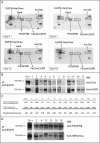
Expression and activation of RTKs. The sample numbers correspond to the cases listed in Table 2. (A) RTK arrays. Equal amounts of total protein extracts from 4 representative cases were incubated with the arrays. The rectangles indicate the presence of activated receptors. (B) EGFR IP/Western blot analysis of 12 representative cases. To confirm the presence of activated EGFR, proteins were immunoprecipitated with a specific antibody and blotted onto a membrane. The anti-pTyr panel identified the phosphorylated EGFR; the anti-EGFR panel indicates the expression of the corresponding receptor. The A431 cell line (American Type Culture Collection, Manassas, VA) was used as the positive control. (C) Densitometric analysis of levels of EGFR activation. The bands representing phosphorylated EGFR and total EGFR levels were quantified, and the ratios between phosphorylated and expressed EGFR were calculated, the ratio relating to the A431 cell line (positive control) was 0.97. The low level of phosphorylation detected in samples #16 and #17 may have been due to methodological problems (non-optimal cryopreservation). (D) Co-IP of EGFR/PDGFRB and EGFR/HER-2/neu. Total protein extracts from 6 EGFR-positive cases were immunoprecipitated with anti-EGFR antibody and run on a gel. The filter was incubated with anti-PDGFRB antibody, then stripped and re-incubated with anti-HER2/neu antibody. Extracts from the 2N5A cell line (immunoprecipitated with anti-PDGFRB) and SKBR3 cell line (immunoprecipitated with anti-HER2/neu) were respectively used as positive controls for PDGFRB and HER2/neu protein. The 2N5A cell line (derived from the NIH3T3 cell line and expressing the COL1-PDGFB fusion that characterized dermatofibrosarcoma protuberans) was kindly provided by Dr. Greco, Fondazione IRCCS Istituto Nazionale dei Tumori; the SKBR3 came from the American Type Culture Collection.
Fig. 3.
Fig. 3.
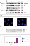
Expression of downstream signaling markers. (A) Western blots showing the presence of phosphorylated/activated and total AKT and ERK1/2. PTEN analyses: (B) FISH. The chromosome 10 probe is labeled with Spectrum Green and the PTEN locus-specific probe with Spectrum Orange. Case 1: monosomic PTEN in 80% of the cells. Case 2: disomic PTEN. (C) Western blotting. PTEN expression in 12 representative monosomic or disomic cases: monosomic cases were Patients 1, 3, 7, 12, and 17. The different electrophoretic mobility observed in Patient 16 may have been due to protein hyper-phosphorylation, which cannot be confirmed by the antibody used because it does not discriminate between phosphorylated and non-phosphorylated PTEN. The A431 and 3962 M cell lines were, respectively, used as positive and negative controls, and anti-actin antibody was used to normalize the results. (D) Real-time PCR in the disomic and monosomic cases. Lymphocyte mRNA was used for calibration purposes.
Fig. 4.
Fig. 4.
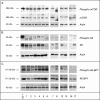
mTOR expression and its downstream effectors. mTOR (panel A), S6 (panel B), and 4E-BP1 (panel C) in the same 12 representative cases analyzed and reported in Figure 3, panel A. Twenty micrograms of total protein extract was loaded for each sample. The A431 cell line was used as the positive control and anti-actin antibody was used to normalize the results.

References

    1. McMaster ML, Goldstein AM, Bromley CM, Ishibe N, Parry DM. Chordoma: incidence and survival patterns in the United States, 1973–1995. Cancer Causes Control. 2001;12:1–11. - PubMed
    1. Mirra JM, Nelson SD, Della Rocca C, Mertens F. Chordoma. In: Fletcher CDM, Unni KK, Mertens F, editors. Pathology and Genetics of Tumors of Soft Tissue and Bone. World Health Organization Classification of Tumors. Lyon: IARC Press; 2002. pp. 316–317.
    1. Chugh R, Tawbi H, Lucas DR, Biermann JS, Schuetze SM, Baker LH. Chordoma: the nonsarcoma primary bone tumor. Oncologist. 2007;12(11):1344–1350. - V体育官网入口 - PubMed
    1. Miozzo M, Dalprà L, Riva P, et al. A tumor suppressor locus in familial and sporadic chordoma maps to 1p36. Int J Cancer. 2000;87(1):68–72. - PubMed (VSports app下载)
    1. Kelley MJ, Korczak JF, Sheridan E, Yang X, Goldstein AM, Parry DM. Familial chordoma, a tumor of notochordal remnants, is linked to chromosome 7q33. Am J Hum Genet. 2001;69(2):454–460. - PMC - PubMed

Publication types

MeSH terms

Substances