Skip to main page content
U.S. flag

An official website of the United States government

Dot gov

The . gov means it’s official. Federal government websites often end in . gov or . mil. Before sharing sensitive information, make sure you’re on a federal government site. VSports app下载.

Https

The site is secure. The https:// ensures that you are connecting to the official website and that any information you provide is encrypted and transmitted securely V体育官网. .

. 2010 Feb 3;11(2):147-60.
doi: 10.1016/j.cmet.2010.01.001.

FoxO1 is a positive regulator of bone formation by favoring protein synthesis and resistance to oxidative stress in osteoblasts

Affiliations

FoxO1 is a positive regulator of bone formation by favoring protein synthesis and resistance to oxidative stress in osteoblasts

Marie-Therese Rached et al. Cell Metab. .

Abstract

Osteoporosis, a disease of low bone mass, is associated with decreased osteoblast numbers and increased levels of oxidative stress within osteoblasts. Since transcription factors of the FoxO family confer stress resistance, we investigated their potential impact on skeletal integrity. Here we employ cell-specific deletion and molecular analyses to show that, among the three FoxO proteins, only FoxO1 is required for proliferation and redox balance in osteoblasts and thereby controls bone formation. FoxO1 regulation of osteoblast proliferation occurs through its interaction with ATF4, a transcription factor regulating amino acid import, as well as through its regulation of a stress-dependent pathway influencing p53 signaling VSports手机版. Accordingly, decreasing oxidative stress levels or increasing protein intake normalizes bone formation and bone mass in mice lacking FoxO1 specifically in osteoblasts. These results identify FoxO1 as a crucial regulator of osteoblast physiology and provide a direct mechanistic link between oxidative stress and the regulation of bone remodeling. .


Figures

Figure 1
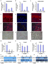
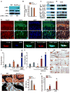
Figure 1. Expression and regulation of FoxO family members in bone
A-C) Expression of all the 3 FoxO genes in primary calvarial osteoblasts, femurs and osteoclasts of WT mice by real-time PCR (n=4 mice/group and duplicates were performed for cell extracts). Bars indicate means ± sem. Expression levels are relative to FoxO1. FoxO1 expression has been considered 1. Mice were 2 months old. D-L) Immunohistochemical localization of FoxO1 in femoral sections of newborn WT mice. (D,and G) Images of bone sections depicting FoxO1 staining at 40× and 100× magnification. (E and H) Sections were counterstained with DAPI. (F and I) FoxO1 and DAPI images were overlaid to visualize nuclear and cytoplasmic localization of FoxO1 at 40× and 100× magnification. Arrows indicate representative cells showing nuclear localization of FoxO1 (purple). (J) DAB staining of FoxO1 in femoral sections. Adjacent sections were stained with FoxO1 and counterstained with (K) eosin or L) hematoxylin (100× magnification). P indicates periosteal surface; E indicates endosteal surface; and, BM indicates bone marrow. The 100× magnification images are obtained from the endosteal surface. M, N and O) Expression analysis of all the 3 FoxO isoforms in murine bones collected from different ages (n=4 mice/group). * p < 0.05 (6 months vs. 2 months); # p < 0.05 (12 months vs. 6 months). Expression levels are relative to FoxO expression at 2 months of age. Expression at 2 months of age has been considered 1. P, Q and R) Activity assessment by phosphorylation status of FoxO1, FoxO3 and FoxO4 in bones collected from mice of different ages. See also Figures S1 and S2.
Figure 2
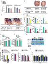
Figure 2. Low bone formation in FoxO1ob-/- mice
A) Bone mineral density (BMD) measured by DEXA in spine and femur of 2 month-old WT, FoxO1ob+/- and FoxO1ob-/- mice (n=10 mice/group). * p < 0.05 vs WT. B) BV/TV, bone volume over trabecular volume; N.Ob/T.Ar, number of osteoblasts per trabecular area; BFR, bone formation rate, of 2 month-old and OcS/BS, osteoclast surface per bone surface in verterbrae of 1 month-old WT and FoxO1ob-/- mice (n=10 mice/group). * p < 0.05 vs WT. C) BV/TV in femoral head and midshaft cortical thickness in the femurs of 2 month-old WT and FoxO1ob-/- mice (n=10 mice/group). * p < 0.05 vs WT. D) BV/TV, BFR and N.Ob/T.Ar in verterbrae of 6 and 12 month-old WT and FoxO1ob-/- mice (n=5-10 mice/group). * p < 0.05 vs WT. E, F) BMD measured by DEXA in spine and femur of 2 month-old WT and FoxO3ob-/- mice (n=10 mice/group). G) Immunoblotting analysis of FoxO4 activity in the bone of WT and FoxO1ob-/- mice. H) RT-PCR analysis of FoxO4 expression in osteoblasts transfected with siRNA oligos for FoxO4 or control (Ctrl), scrambled oligos. * p < 0.05 vs control (Ctrl) siRNA and Ctrl siRNA in OM. OM denotes Osteogenic Medium. I) Proliferation in cultures of osteoblasts transfected with siRNA oligos for FoxO4 or control (Ctrl), scrambled oligos. OM denotes Osteogenic Medium. J) Real-time PCR (RT-PCR) analysis of CyclinD1, D2 and p27Kip1 expression in osteoblasts with silenced FoxO4. K) RT-PCR analysis of osteoblast differentiation markers in osteoblasts with silenced FoxO4. In all panels values are means ± sem.
Figure 3
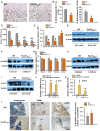
Figure 3. Decreased osteoblast proliferation in bones of FoxO1Ob-/- mice
A) Sections of femurs from d5.5 WT and FoxO1ob-/- pups stained with BrdU (Image, 40× magnification) B) Osteoblast proliferation expressed as number of BrdU stained osteoblasts per Trabecular Area (T.Ar.) and per Bone Perimeter (B.Pm) (n=4 mice/group). * p < 0.05 vs WT. C) RT-PCR analysis of FoxO1, FoxO3 and FoxO4 expression in the bone of WT and FoxO1ob-/- mice at indicated ages. d denotes day and M demotes months (n=3 mice per group). * p < 0.05 vs WT; # p < 0.05 vs WT at d5.5; $ p < 0.05 vs WT at 1M. D) RT-PCR analysis of CyclinD1, D2 and p27Kip1 expression in femurs of WT and FoxO1ob-/- mice at 2 months of age (n=6 mice/group). E and F) Immunoblotting of cyclins D1 and D2 in bones of WT and FoxO1ob -/- mice. G) RT-PCR analysis of Runx2, BSP, Osterix and Col1a1 expression in bones of WT and FoxO1ob -/- mice (n=6 mice/group). * p < 0.05 vs WT. H and I) Immunoblotting of Runx2 and Type I collagen in bones of WT and FoxO1ob -/- mice. J and K) RT-PCR analysis of Runx2 and Alkaline phosphatase (ALP) expression in cultured primary calvarial osteoblasts of WT and FoxO1ob-/- mice (n=3) treated with vehicle or osteogenic medium. * p < 0.05 vs vehicle L) Apoptosis in bone sections from the femurs of 3 month-old WT and FoxO1ob-/- mice (n=4 mice per group). Arrows indicate osteoblasts (left panel) or apoptotic osteoblasts (right panel). Control indicates TUNEL staining of a bone section treated with DNAse. Magnifications are 40× unless otherwise stated. The 100× magnification panel shows an example of a group of apoptotic osteoblasts. In all panels values are means ± sem.
Figure 4
Figure 4. N-acetyl L-cysteine (NAC) rescues the increased oxidative stress and the bone phenotype of FoxO1ob-/- mice
A) SOD2 activity in femurs of WT and FoxO1ob-/- mice (u/mg) (n=6 mice/group). * p < 0.05 vs WT. B) RT-PCR analysis of FasL and Gadd45 expression in femurs of WT and FoxO1ob-/- mice (n=5 mice/group). * p < 0.05 vs WT. C) Glutathione (GSH) levels in bones of WT and FoxO1ob-/- mice (n=5 mice/group). * p < 0.05 vs WT. D) Flow cytometry analysis of reactive oxygen species (ROS) levels in osteoblasts from bone marrow cells of WT and FoxO1ob-/- mice (n=5 mice/group). * p < 0.05 vs WT. E) Lipid peroxidation levels in bones of WT and FoxO1ob-/- mice (n=5 mice/group). * p < 0.05 vs WT. F and G) p53 and p66shc activity measured by immunoblotting in bones of WT and FoxO1ob-/- mice (n=2 mice/group). H) Densitometric analysis of the ratio of p-p53 / p53. ND denotes not determined. I and J) RT-PCR analysis of p19ARF and p16 expression in bones of WT and FoxO1ob-/- mice (n=5 mice/group). * p < 0.05 vs WT. K) RT-PCR analysis of p19ARF and p16 expression in bones of WT and FoxO3ob-/- mice (n=5 mice/group). L) GSH levels in osteoblasts of vehicle- or NAC-treated WT and FoxO1ob-/- mice (n=5 mice/group). * p < 0.05 vs WT vehicle and WT NAC. M and N) RT-PCR analysis of p19ARF and p16 expression in bones of vehicle- or NAC-treated WT and FoxO1ob-/- mice (n=5 mice/group). * p < 0.05 vs WT vehicle; # p < 0.05 vs WT vs FoxO1ob-/- vehicle. O) BV/TV, N.Ob./T.Ar and BFR in spines of vehicle or NAC-treated WT and FoxO1ob-/- mice (n=5 mice/group). * P < 0.05 (FoxO1ob-/- vehicle vs. WT); # P < 0.05 (FoxO1ob-/- NAC vs FoxO1ob-/- vehicle). In all panels, except I and J where mice were 5.5 days of age, animals were 3 months of age; and, values are means ± sem. See also Figures S3 and S4.
Figure 5
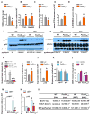
Figure 5. Altered amino acid metabolism in osteoblasts of FoxO1ob-/- mice
A) Immunoblotting analysis of the phosphorylation status of eIF2α̣ in bone from WT and FoxO1ob-/- mice (n=5 mice/group). B) RT-PCR analysis of ATF4 expression in bone from WT and FoxO1ob-/- mice (n=5 mice/group). C and D) Immumoprecipitation (IP) and immunoblotting of FoxO1 and ATF4 in nuclear extracts from primary osteoblasts and bones of WT mice. E) Immunohistochemical localization of FoxO1 and ATF4 in femoral sections of newborn, WT mice. Images of bone sections depicting FoxO1, ATF4, DAPI and combination of ATF4 with DAPI or FoxO1 with ATF4 stainings. The top 5 panels show 40× and the 5 lower panels show 100× magnifications. P indicates periosteal surface; E indicates endosteal surface; and, BM indicates bone marrow. The 100× magnification images are obtained from the endosteal surface. F) Immunohistochemical localization of FoxO1 and ATF4 in primary osteoblasts. Single cell images at 100× magnification show staining with the indicated antibodies. G) Co-transfection of FoxO1, ATF4 and FoxO-Luc reporter construct in COS-7 cells. EV denotes empty vector. Results are presented as fold induction over EV. (EV=1). * p < 0.05 vs.FoxO-luc; # P < 0.05 vs FoxO1/FoxO-luc. H) Co-transfection of FoxO1, ATF4 and OG2-Luc reporter construct in COS-7 cells. EV denotes empty vector. Results are presented as fold induction over EV. (EV=1). * P < 0.05 vs. OG2-luc; # P < 0.05 vs ATF4/OG2-Luc and vs FoxO1/OG2-luc). I) Von Gieson staining indicating collagen content (stained red) on vertebral sections from 2, 6 and 12 month-old WT and FoxO1ob-/- mice. Representative results from n = 5 mice per group. J) Osteoid surface by Von Kossa staining in vertebral sections from 1 month-old WT and FoxO1ob-/- mice (n= 6 mice per group). OS and BS denote osteoid surface and bone surface, respectively. Arrows indicate osteoid deposition. * p < 0.05 vs WT. K) RT-PCR analysis of p19ARF and p16 expression in primary osteoblasts derived from Atf4-/- mice. * p < 0.05 vs WT. In all panels values are means ± sem.
Figure 6
Figure 6. High protein diet (HPD) rescues the bone phenotype of FoxO1ob-/- mice
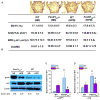
A) BV/TV, N.Ob/T.Ar., BFR and osteoclast surface in the verterbrae of WT and FoxO1ob-/- on normal (ND) or HPD (n=5 mice/group). * P < 0.05 (FoxO1ob-/- ND vs. WT ND); # P < 0.05 (FoxO1ob-/- HPD vs FoxO1ob-/- ND). The OcS/BS data shown for WT and FoxO1ob-/- mice on normal diet, are identical to those shown in Figure 2B. B) Immunoblotting of phospho-p53 in bones of WT and FoxO1ob-/- on ND or HPD. C and D) RT-PCR analysis of p19ARF and p16 expression in bones of WT and FoxO1ob-/- on ND or HPD (n=5 mice/group). * P < 0.05 (FoxO1ob-/- vs. WT); # P < 0.05 (FoxO1ob-/- HPD vs FoxO1ob-/- ND). In all panels, mice were 1 month of age.
Figure 7
Figure 7. Model depicting the mechanism of FoxO1 action in osteoblasts
Under physiological levels of stress, FoxO1 shuttles between the nucleus and the cytoplasm. In the nucleus, FoxO1 interacts with ATF4. This interaction promotes the transcriptional activity of FoxO1 and is required for amino acid import and protein synthesis. Normal protein synthesis allows FoxO1 to orchestrate an anti-oxidant defense mechanism that maintains redox balance by suppressing expression of p19ARF and p16 and downstream activation of their target protein p53. Transcriptional repression of the p19ARF/p16/p53 pathway prevents cell cycle arrest in osteoblasts and maintains their normal proliferation and bone homeostasis

References

    1. Accili D, Arden KC. FoxOs at the crossroads of cellular metabolism, differentiation, and transformation. Cell. 2004;117:421–426. - PubMed
    1. Almeida M, Han L, Martin-Millan M, Plotkin LI, Stewart SA, Roberson PK, Kousteni S, O'Brien CA, Bellido T, Parfitt AM, Weinstein RS, Jilka RL, Manolagas SC. Skeletal involution by age-associated oxidative stress and its acceleration by loss of sex steroids. J Biol Chem. 2007;282:27285–27297. - PMC - PubMed
    1. Arden KC. Multiple roles of FOXO transcription factors in mammalian cells point to multiple roles in cancer. Exp Gerontol. 2006;41:709–717. - PubMed (V体育官网入口)
    1. Bai XC, Lu D, Bai J, Zheng H, Ke ZY, Li XM, Luo SQ. Oxidative stress inhibits osteoblastic differentiation of bone cells by ERK and NF-kappaB. Biochem Biophys Res Commun. 2004;314:197–207. - "VSports在线直播" PubMed
    1. Bouxsein ML, Melton LJ, III, Riggs BL, Muller J, Atkinson EJ, Oberg AL, Robb RA, Camp JJ, Rouleau PA, McCollough CH, Khosla S. Age- and sex-specific differences in the factor of risk for vertebral fracture: a population-based study using QCT. J Bone Miner Res. 2006;21:1475–1482. - PubMed

Publication types

Substances

"V体育平台登录" LinkOut - more resources