Skip to main page content
U.S. flag

An official website of the United States government

Dot gov

The VSports app下载. gov means it’s official. Federal government websites often end in . gov or . mil. Before sharing sensitive information, make sure you’re on a federal government site. .

Https

The site is secure. The https:// ensures that you are connecting to the official website and that any information you provide is encrypted and transmitted securely. V体育官网.

. 2010 Jul;130(3):363-73.
doi: 10.1111/j.1365-2567.2009.03236.x. Epub 2010 Feb 5.

Enhancement of effector CD8+ T-cell function by tumour-associated B7-H3 and modulation of its counter-receptor triggering receptor expressed on myeloid cell-like transcript 2 at tumour sites

Affiliations

Enhancement of effector CD8+ T-cell function by tumour-associated B7-H3 and modulation of its counter-receptor triggering receptor expressed on myeloid cell-like transcript 2 at tumour sites

V体育2025版 - Hiroko Kobori et al. Immunology. 2010 Jul.

Abstract

B7-H3 is a B7-family co-stimulatory molecule and is broadly expressed on various tissues and immune cells. Transduction of B7-H3 into some tumours enhances anti-tumour responses. We have recently found that a triggering receptor expressed on myeloid cell-like transcript 2 (TLT-2) is a receptor for B7-H3. Here, we examined the roles of tumour-associated B7-H3 and the involvement of TLT-2 in anti-tumour immunity. Ovalbumin (OVA)(257-264)-specific OT-I CD8(+) T cells exhibited higher cytotoxicity against B7-H3-transduced OVA-expressing tumour cells (B7-H3/E. G7) in vitro and selectively eliminated B7-H3/E. G7 cells in vivo. The presence of B7-H3 on target cells efficiently augmented CD8(+) T-cell-mediated cytotoxicity against alloantigen or OVA, whereas the presence of B7-H3 in the priming phase did not affect the induced cytotoxicity. B7-H3 transduction into five tumour cell lines efficiently reduced their tumorigenicity and regressed growth. Treatment with either anti-B7-H3 or anti-TLT-2 monoclonal antibody accelerated growth of a tumour that expressed endogenous B7-H3, suggesting a co-stimulatory role of the B7-H3-TLT-2 pathway. The TLT-2 was preferentially expressed on CD8(+) T cells in regional lymph nodes, but was down-regulated in tumour-infiltrating CD8(+) T cells VSports手机版. Transduction of TLT-2 into OT-I CD8(+) T cells enhanced antigen-specific cytotoxicity against both parental and B7-H3-transduced tumour cells. Our results suggest that tumour-associated B7-H3 directly augments CD8(+) T-cell effector function, possibly by ligation of TLT-2 on tumour-infiltrating CD8(+) T cells at the local tumour site. .


Figures
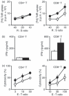
Figure 1
Figure 1
Enhancement of CD8+ T-cell effector function by B7-H3. CD4+ or CD8+ T cells were co-cultured with either P815 (white circles or columns) or B7-H3/P815 (black circles or columns) in the presence of a sub-optimal dose of anti-CD3 monoclonal antibody at the indicated responder : stimulator (R : S) or effector : target (E : T) ratios. Proliferative responses for 3 days (a), interferon-γ (IFN-γ) production for 4 days culture (b) and cytotoxicity for 6 hr (c) were measured as described in the Materials and methods. Values shown are the means ± SD from triplicate cultures. The data are representative of three independent experiments.
Figure 2
Figure 2
Selective lysis in B7-H3-transduced tumours. Freshly isolated OT-I CD8+ T cells (a), in vitro-sensitized OT-I CD8+ T cells (b) were used as effector cells. Cytotoxicity against parental E.G7 (white circles) or B7-H3/E.G7 (black circles) was measured. Values shown are means ± SD. (c) The mixture of [5-(and-6)-(((4-chloromethyl)benzoyl)amino)] tetramethylrhodamine (CMTMR) -labelled E.G7 and carboxyfluorescein diacetate succinimidyl ester (CFSE) -labelled E.G7 (1 : 2, A-mix) or CMTMR-labelled E.G7 and CFSE-labelled B7-H3/E.G7 (1 : 2, B-mix) was injected into the peritoneal cavity of OT-I mice. After 24 hr, peritoneal exudate cells (PEC) were analysed by flow cytometry. The same number (5 × 10³ cells) of CMTMR+ cells was acquired, and data are displayed as dotted plots with CFSE (x-axis) and CMTMR (y-axis) intensity. Representative profiles from two independent experiments are shown. Values in the profiles are the mean positive percentages ± SD from each group of three mice. In the right panel, the values shown are the CFSE : CMTMR ratios of the isolated PEC after the injection of A and B mix (the mean ± SD).
Figure 3
Figure 3
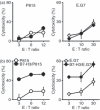
Enhanced cytotoxic T lymphocytes (CTL) against alloantigen and ovalbumin (OVA) by B7-H3-expressing target cells. (a) B6 (left panel) and OT-I (right panel) mice were sensitized in vivo with allogeneic tumour cells (P815 or B7-H3/P815) and OVA-expressing tumour cells (E.G7 or B7-H3/E.G7), respectively. After the sensitization, cytotoxicity against respective parental tumour cells was measured using peritoneal exudate cells as effector cells. (b) B6 (left panel) and OT-I (right panel) mice were sensitized in vivo with P815 and E.G7 cells, respectively. After the sensitization, cytotoxicity against respective parental and B7-H3-transfected tumour cells was measured. Values shown are the means ± SD. The data are representative of three independent experiments.
Figure 4
Figure 4
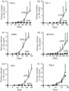
Reduced tumorigenicity by B7-H3 transduction. Parental (white circles) and B7-H3-transduced (black circles) tumour cells (a, P815; b, EL4; c, J558L; d, SCCVII; e, B16) were subcutaneously injected into syngeneic mice and tumour volume was monitored. In (f ), parental and B7-H3-transduced P815 tumour cells were injected into BALB/c nude mice as described above. The mean tumour volume ± SD (mm3) was determined in each group of five or six mice, and the data are representative of two independent experiments. The final tumour rejected ratios are shown in brackets.
Figure 5
Figure 5
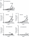
Requirements for B7-H3-induced anti-tumour immunity (a) Mice were pre-treated with control rat immunoglobulin G (IgG; white squares), anti-CD4 (black triangles), anti-CD8 (black diamonds), or both anti-CD4 and anti-CD8 (black circles) monoclonal antibodies (mAbs), and then B7-H3/SCCVII tumour cells were inoculated and tumour volume was monitored. The mean tumour volume ± SD (mm3) was determined. The final tumour rejected ratios are shown in brackets. The data shown are representative of two independent experiments. (b) and (c) SCCVII or B7-H3/SCCVII tumour-inoculated mice received intraperitoneal injections of control rat IgG (white circles) or anti-B7-H3 mAb (black circles) (in b), and control rat IgM (white diamonds) or anti-TLT-2 mAb (black diamonds) (in c). The tumour volume was monitored. The mean tumour volume ± SD (mm3) was determined in each group of 11 mice from two independent experiments.
Figure 6
Figure 6
Modulation of TLT-2 expression on CD8+ T cells and enhanced cytotoxicity by TLT-2. (a) Lymph node cells from naive mice and regional lymph node cells and tumour-infiltrating lymphocytes (TILs) were obtained from SCCVII or B7-H3/SCCVII tumour-inoculated mice. Cells were stained with fluorescein isothiocyanate-conjugated (FITC-) anti-CD45, peridinin chlorophyll protein Cychrome 5.5-conjugated (PerCP-Cy5.5-) anti-CD3, and either phycoerythrin-conjugated (PE-) anti-CD4 or anti-CD8 mAbs, and biotinylated anti-TLT-2 mAb, followed by allophycocyanin (APC) -streptavidin or with the appropriate fluorochrome-conjugated control immunoglobulins and were then analysed by flow cytometry. An electronic gate was placed on CD45+ CD3+ CD8+ lymphocytes and TLT-2 expression is displayed as histograms with the control histograms nearest the ordinate (shaded). The data are representative of two independent experiments. (b) TILs from B7-H3/SCCVII-inoculated mice were stained with FITC-anti-CD45, PerCP-Cy5.5-anti-CD8, PE-anti-CD69 mAbs and biotinylated anti-TLT-2 mAb, followed by APC-streptavidin. CD8+ T cells from naive OT-I mice were labelled with carboxyfluorescein diacetate succinimidyl ester (CFSE) and co-cultured with E.G7 or B7-H3/E.G7 cells for 24 hr. The harvested cells were stained with either PE-anti-CD69 or anti-CD25, and PerCP-Cy5.5-anti-CD8 mAbs and biotinylated anti-TLT-2 mAb, followed by APC-streptavidin. The cells were analysed by flow cytometry. An electronic gate was first placed on CFSE+ CD8+ lymphocytes and then the secondary gates were set on CD69- or CD25-positive and negative cells. TLT-2 expression is displayed as histograms with the control histograms nearest the ordinate (shaded). The values of upper right are the mean fluorescence intensity (MFI) of TLT-2 stained cells. The data are representative of two independent experiments. (c) CD8+ T cells from naive B6 mice were stimulated with immobilized anti-CD3 mAb in the presence of the indicated cytokines for 3 days. The cells were harvested and stained with FITC-anti-CD8, PerCP-Cy5.5-anti-CD3 mAbs and biotinylated anti-TLT-2 mAb, followed by APC-streptavidin and analysed by flow cytometry. The data are representative of two independent experiments. The values of upper right are the MFI of TLT-2 stained cells. (d) Mock- or TLT-2-transduced OT-I CD8+ T cells were stained with biotinylated anti-TLT-2 mAb, followed by PE-streptavidin, and analysed by flow cytometry. The data are displayed as histograms with the control histograms nearest the ordinate (shaded). Mock-transduced (white circles) or TLT-2-transduced (black circles) OT-I CD8+ T cells were used as effector cells. Cytotoxicity against E.G7 or B7-H3/E.G7 cells was measured. Values shown are the means ± SD. The data are representative of two independent experiments.
Figure 7
Figure 7
Abundant tumour-infiltrating lymphocytes (TIL) in B7-H3/SCCVII-inoculated sites. The abdominal skin of SCCVII or B7-H3/SCCVII tumour (1 × 10⁵ cells) inoculated sites was surgically resected after 7 days. The formalin-fixed, paraffin-embedded tissue sections were stained with haematoxylin & eosin. Representative images of the tumour mass near the epithelium from three individuals are shown. Lower images show the selected field of the upper images at higher magnification. Scale bars = 100 μm.

References

    1. Chapoval AI, Ni J, Lau JS, et al. B7-H3: a costimulatory molecule for T cell activation and IFN-γ production. Nat Immunol. 2001;2:269–74. - PubMed
    1. Sun JC, Bevan MJ. Defective CD8 T cell memory following acute infection without CD4 T cell help. Science. 2003;300:339–42. - PMC - PubMed
    1. Zhang GB, Dong QM, Xu Y, Yu GH, Zhang XG. B7-H3: another molecule marker for Mo-DCs? Cell Mol Immunol. 2005;2:307–11. - PubMed
    1. Kim J, Myers AC, Chen L, et al. Constitutive and inducible expression of b7 family of ligands by human airway epithelial cells. Am J Respir Cell Mol Biol. 2005;33:280–9. - PMC - PubMed
    1. Saatian B, Yu XY, Lane AP, Doyle T, Casolaro V, Spannhake EW. Expression of genes for B7-H3 and other T cell ligands by nasal epithelial cells during differentiation and activation. Am J Physiol Lung Cell Mol Physiol. 2004;287:L217–25. - PubMed

Publication types

VSports注册入口 - MeSH terms

LinkOut - more resources