Skip to main page content (V体育ios版)
U.S. flag

An official website of the United States government

Dot gov

The . gov means it’s official. Federal government websites often end in . gov or . mil. Before sharing sensitive information, make sure you’re on a federal government site VSports app下载. .

Https

The site is secure. The https:// ensures that you are connecting to the official website and that any information you provide is encrypted and transmitted securely V体育官网. .

. 2010 Mar;11(3):207-15.
doi: 10.1038/ni.1839. Epub 2010 Jan 17.

V体育官网入口 - Regulation of hematopoietic stem cell differentiation by a single ubiquitin ligase-substrate complex

Affiliations

Regulation of hematopoietic stem cell differentiation by a single ubiquitin ligase-substrate complex

Linsey Reavie et al. Nat Immunol. 2010 Mar.

Abstract

Hematopoietic stem cell (HSC) differentiation is regulated by cell-intrinsic and cell-extrinsic cues. In addition to transcriptional regulation, post-translational regulation may also control HSC differentiation. To test this hypothesis, we visualized the ubiquitin-regulated protein stability of a single transcription factor, c-Myc. The stability of c-Myc protein was indicative of HSC quiescence, and c-Myc protein abundance was controlled by the ubiquitin ligase Fbw7. Fine changes in the stability of c-Myc protein regulated the HSC gene-expression signature. Using whole-genome genomic approaches, we identified specific regulators of HSC function directly controlled by c-Myc binding; however, adult HSCs and embryonic stem cells sensed and interpreted c-Myc-regulated gene expression in distinct ways. Our studies show that a ubiquitin ligase-substrate pair can orchestrate the molecular program of HSC differentiation VSports手机版. .


Figures
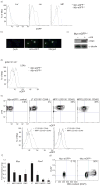
Figure 1
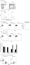
Figure 1. Visualization of c-Myc protein abundance in early hematopoiesis
a) Levels of c-Myc-eGFP expression during early hematopoiesis in Lineage+, Lineage, LSK (Lineage, c-Kit+, Sca1+) and MP (lineage, c-Kit+, Sca1) BM subsets. n=6 mice b) Immuno-fluorescence tracing of nuclear c-Myc-eGFP expression in Lineage bone marrow progenitors. c) Western blot demonstrating presence of c-Myc protein in c-Kit+ and absence in c-Kit subset of adult bone marrow. d) Overlay histogram depicting c-Myc-eGFP protein expression within LSK cells. e) c-Myc-eGFP expression in eGFP−/−control, LT-HSC and MPP (1, 2) populations. f) qRT-PCR quantification of Myc and Fbw7 mRNA expression during HSC differentiation. g) Levels of c-Myc-eGFP protein correlate with the cell cycle status of LSK cells. Sorted c-Myc-eGFPHi or c-Myc-eGFPLo LSK cells were analyzed using Ki-67 and DAPI staining. Plots are a representation of at least 3 independent experiments.
Figure 2
Figure 2. c-Myc protein levels correlate with loss of HSC self-renewal
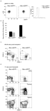
a) FACS-dependent separation of c-Myc-eGFPHi and c-Myc-eGFPLo LSK populations. b) CFU in vitro cultures using sorted c-Myc-eGFPHi and c-Myc-eGFPLo cells. In black: First plating of LSKs, in grey: second plating. Error bars indicate standard deviation (Std) (n=3 mice in 3 independent experiments). c) Peripheral blood chimerism in competitive reconstitution assays at 25 weeks post transplant (n=3 mice). d) Relative chimerism of sorted c-Myc-eGFPHi and c-Myc-eGFPLo LSKs (CD45.2) in the bone marrow (LSK subset), thymus and spleen (d) 17 weeks post transplantation (n=3 mice).
Figure 3
Figure 3. c-Myc protein stability in HSC is controlled by the ubiquitin ligase Fbw7
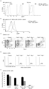
a) FACS analysis showing detection of c-Myc-eGFP in different progenitor populations. LSK, MP and thymic DN (CD48) subsets are shown. b) FACS analysis showing stabilization of c-Myc-eGFP protein in LT-HSC subset (LSK, CD150+, CD48) upon Fbw7 deletion c-d) Deletion of a single Myc allele significantly rescues the Fbw7−/− HSC phenotype. FACS profiles of bone marrow LSKs (c), cell cycle status of LSKs (d) and methylcellulose assays (CD150+ LSKs) (e) are shown. Black: first plating, Grey: second plating. Error bars indicate standard deviation (Std) (n=5 mice). Plots are a representation of at least 3 independent experiments.
Figure 4
Figure 4. The role of the c-Myc:Fbw7 interaction in fetal liver stem and progenitor cells
a) FACS staining defining LSK cells in e.d.14.5 fetal liver and adult (6wk old) bone marrow. b) Cell cycle status of fetal and adult LSK cells. c) Levels of c-Myc-eGFP protein expression in fetal and adult LSK cells. The overlay histogram shows induction of c-Myc protein expression in fetal LSKs. d) Levels of c-Myc-eGFP protein expression in CD150+ LSKs. e) Methylcellulose culture using the indicated cell populations purified from the fetal liver (fetal) or the bone marrow (adult). Black: first plating, Grey: second plating. Error bars indicate standard deviation (Std) (n= 6 mice). f) c-Myc-eGFPLo but not c-Myc-eGFPHi expressing fetal liver LSK subsets show chimerism in the peripheral blood 20wks post transplant in competitive reconstitution assays. CD45.2+ cells are donor-derived cells in the peripheral blood (n= 6 mice). Plots are a representation of at least 3 independent experiments.
Figure 5
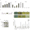
Figure 5. c-Myc protein abundance directly controls the molecular program of stem cell differentiation, cell cycle entry and self-renewal
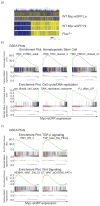
a) Heat map demonstrating relative gene expression signatures of c-Myc-eGFPHi and c-Myc-eGFPLo subsets. A direct comparison to the relative gene expression in Fbw7−/− LSKs is also shown. b) Representative GSEA profiles showing positive correlation of the c-Myc-eGFPLo signature to stem cell gene sets and negative correlation to cell cycle and DNA replication gene sets. c) Representative GSEA profiles showing a positive correlation between both TGF-β and Wnt signaling pathways with c-Myc-eGFPLo gene expression data set.
Figure 6
Figure 6. Genes over-expressed in c-Myc-EGFPHi cells are directly bound by the c-Myc transcription factor
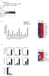
a) GSEA analysis showing a positive correlation of c-Myc-eGFPHi gene expression profiles to a direct c-Myc target Chip-on-chip dataset. b) ChIP assay and Heat map of selected genes that are over-expressed in the c-Myc-eGFPHi cells. Red color: gene upregulation, Blue color: gene down-regulation. Two individual chromatin IPs using T-ALL cell line genomic DNA are shown. c) ChIP assay of selected genes using genomic DNA from purified Lineage c-KiteGFP+ bone marrow progenitor cells. Also, Heat Map showing over expression of selected genes in the c-Myc-eGFPHi LSK subset. Error bars indicate standard deviation (std) from 3 independent experiments.
Figure 7
Figure 7. Patterns of Fbw7 and c-Myc expression in mouse embryonic stem cells
a) mRNA transcript expression levels of Fbw7 (black), Myc (dark grey) and Nanog (light grey) in self-renewing (+LIF) and differentiating (−LIF, +RA) mouse ESC. Different days of differentiation (d0–6) are shown), b) Schematic diagram of Fbw7 gene-trap cassette. c) LacZ staining in Murine ESCs containing the Fbw7 gene trap cassette depicting upregulation of Fbw7 expression as Murine ESCs differentiate via the removal of LIF and the addition of Retinoic Acid (RA). d) Western blot showing stabilization of both phospho- (T58) and total-c-Myc in self-renewing and differentiating Murine ESCs treated with 20uM of proteosome inhibitor MG-132. e) ChIP assay was carried out using the specific regulators shown in Fig 6 revealing substantial enrichment upon c-Myc immunoprecipitation in Murine ESCs. Error bars indicate standard deviation (std) from 3 independent experiments.
Figure 8
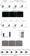
Figure 8. Fbw7 is dispensable for the self-renewal of Murine ESCs
a) Inhibition of proteasomal degradation via MG-132 treatment resulted in accumulation of c-Myc protein in MyceGFP/ Murine ESCs. b) Visualization of c-Myc-eGFP in W4 and MyceGFP/ Murine ESCs with and without treatment with MG-132. GFP (green), nuclear staining (DAPI) c) Knock-down of Fbw7 using siRNA in c-MyceGFP/ ESCs resulted in accumulation of c-Myc protein (increase in eGFP levels) while siRNA targeting both Myc and GFP showed a reduction in c-Myc-eGFP levels as assessed by FACs analysis. d) qRT-PCR of Fbw7 mRNA expression in ESCs expressing shRNAs against Fbw7 and Nanog. A non silencing shRNA is used as a control. e) Western blot depicting accumulation of both phospho (T58)-and total-c-Myc protein upon shRNA mediated knock down of Fbw7 when compared to a non-silencing shRNA control. f) Alkaline phosphatase staining in Murine ESCs showed no difference in differentiation capacity upon shRNA mediated knock-down of Fbw7 when compared to non-silencing control while silencing of Nanog resulted in complete differentiation. g) siRNA knock-down of Fbw7 in a Nanog-eGFP reporter ESC line resulted in no difference in Nanog-eGFP expression levels when compared to non-silencing control. Knock-down of Oct4 resulted in a reduction of Nanog-eGFP expression levels (marking differentiation). Plots are a representation of at least 3 independent experiments

Comment in

References

    1. Adams GB, Scadden DT. The hematopoietic stem cell in its place. Nat Immunol. 2006;7:333–337. - PubMed
    1. Kiel MJ, Morrison SJ. Uncertainty in the niches that maintain haematopoietic stem cells. Nat Rev Immunol. 2008;8:290–301. - PubMed
    1. Kondo M, et al. Biology of hematopoietic stem cells and progenitors: implications for clinical application. Annu Rev Immunol. 2003;21:759–806. - PubMed
    1. Yoshida T, et al. The role of the chromatin remodeler Mi-2beta in hematopoietic stem cell self-renewal and multilineage differentiation. Genes Dev. 2008;22:1174–1189. - PMC - PubMed
    1. Moore KA, Lemischka IR. Stem cells and their niches. Science. 2006;311:1880–1885. - PubMed

Publication types

MeSH terms

"VSports最新版本" Substances