Skip to main page content (V体育安卓版)
U.S. flag

An official website of the United States government

Dot gov

The . gov means it’s official. Federal government websites often end in . gov or VSports app下载. mil. Before sharing sensitive information, make sure you’re on a federal government site. .

Https

The site is secure. The https:// ensures that you are connecting to the official website and that any information you provide is encrypted and transmitted securely V体育官网. .

. 2010 Jan 12;107(2):827-31.
doi: 10.1073/pnas.0908126107. Epub 2009 Dec 22.

Ablation of SLP-76 signaling after T cell priming generates memory CD4 T cells impaired in steady-state and cytokine-driven homeostasis

Affiliations

Ablation of SLP-76 signaling after T cell priming generates memory CD4 T cells impaired in steady-state and cytokine-driven homeostasis

Nicholas D Bushar et al. Proc Natl Acad Sci U S A. .

Abstract

The intracellular signaling mechanisms regulating the generation and long-term persistence of memory T cells in vivo remain unclear. In this study, we used mouse models with conditional deletion of the key T cell receptor (TCR)-coupled adaptor molecule SH2-domain-containing phosphoprotein of 76 kDa (SLP-76), to analyze signaling mechanisms for memory CD4 T cell generation, maintenance, and homeostasis. We found that ablation of SLP-76 expression after T cell priming did not inhibit generation of phenotypic effector or memory CD4 T cells; however, the resultant SLP-76-deficient memory CD4 T cells could not produce recall cytokines in response to TCR-mediated stimulation and showed decreased persistence in vivo VSports手机版. In addition, SLP-76-deficient memory CD4 T cells exhibited reduced steady-state homeostasis and were impaired in their ability to homeostatically expand in vivo in response to the gamma(c) cytokine IL-7, despite intact proximal signaling through the IL-7R-coupled JAK3/STAT5 pathway. Direct in vivo deletion of SLP-76 in polyclonal memory CD4 T cells likewise led to impaired steady-state homeostasis as well as impaired homeostatic responses to IL-7. Our findings demonstrate a dominant role for SLP-76-dependent TCR signals in regulating turnover and perpetuation of memory CD4 T cells and their responses to homeostatic cytokines, with implications for the selective survival of memory CD4 T cells following pathogen exposure, vaccination, and aging. .


Conflict of interest statement

The authors declare no conflict of interest.

"V体育ios版" Figures

Fig. 1.
Fig. 1.
Downmodulation of SLP-76 expression after T cell activation using a conditional knockout mouse model. (A) Kinetics of SLP-76 knockdown. Splenic CD4+ T cells from SLP-76F/nullR26Ryfp (F/null) and SLP-76F/+R26Ryfp (F/+) mice were activated with anti-CD3/anti-CD28 antibodies and cultured with TATCre protein, and SLP-76 expression was determined by flow cytometry 6–24 h later. Histograms show intracellular SLP-76 expression gated on total CD4+ T cells or YFP+CD4+ T cells (for 19 and 24 h). Shaded histograms represent isotype controls. (B) SLP-76 deletion occurs in the YFP+ fraction of cKOTATCre cells. (Left) Western blot of SLP-76 and LAT expression in CD4 T cells from F/null and F/+ mice before stimulation (Prestim) and sorted, CD4+YFP+ cKO or cHET cells after TATCre. The line separates the noncontiguous lanes. (Right) Flow cytometric analysis of YFP versus SLP-76 expression in cKOTATCre and cHETTATCre CD4 T cells analyzed 48 h post-TATCre administration, gated on CD4+ T cells, representative of seven experiments. (C) Surface expression of CD44, CD25, and IL-7Rα on cKOTATCre (Upper) and cHETTATCre (Lower) primed cells gated on CD4+YFP+cells. Results are representative of six experiments. (D) Intracellular phospho-PLC-γ1 expression of cKOTATCre and cHETTATCre primed CD4 T cells gated on CD4+YFP+cells, with shaded histograms denoting control. Results are representative of three experiments.
Fig. 2.
Fig. 2.
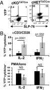
In vivo transfer of primed SLP-76-deficient CD4 T cells. CD4 T cells from cKO and cHET mice were primed as in Fig.1, transferred into RAG2−/− adoptive hosts, and recovered 2 weeks posttransfer. (A) SLP-76 and YFP expression of persisting cKOTATCre and cHETTATCre CD4 T cells, representative of three independent experiments. (B) IL-2 and IFN-γ production by persisting cKOTATCre and cHETTATCre CD4 T cells stimulated for 6 h with anti-CD3/anti-CD28 antibodies (Upper) or PMA/ionomycin (Lower) as determined by intracellular cytokine staining. Results are expressed as mean percentage of cytokine+YFP+ CD4 T cells (n = 6), from two experiments.
Fig. 3.
Fig. 3.
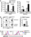
Reduced persistence and homeostasis of SLP-76–deficient memory CD4 T cells. CD4 T cells from cKO and cHET mice were primed in vitro for 72 h, TATCre treated, and transferred into B6.CD45.1 hosts. Persisting cKOTATCre and cHETTATCre memory CD4 T cells were harvested from spleen and lymph nodes 1–5 weeks posttransfer. (A) Surface expression of CD44, CD25, IL-7Rα, and TCRβ gated on CD45.2+CD4+YFP+ cells of cKOTATCre and cHETTATCre memory CD4 T cells 5 weeks posttransfer. Shaded histograms are controls. Results are representative of three experiments (n = 5 mice per group). (B) Persistence of cKOTATCre and cHETTATCre CD4+YFP+ memory CD4 T cells in adoptive hosts calculated by dividing the average number of YFP+ memory T cells at >5 weeks posttransfer by the average number of YFP+ cells at day 6 posttransfer, with n = 12–14 mice for cKOTATCre and n = 9–10 mice for cHETTATCre. (C) Flow cytometry plots of BrdU incorporation in cKOTATCre and cHETTATcre memory CD4 T cells gated on YFP+CD45.2+ CD4+ cells.
Fig. 4.
Fig. 4.
Impaired homeostasis of SLP-76–deficient memory CD4 T cells to IL-7/anti-IL-7 complexes. (A) (Left) Absolute numbers of host splenocytes in untreated and IL-7/M25-treated hosts of cKOTATCre and cHETTATCre memory cells. (Right) Absolute numbers of CD45.2+ YFP+CD4+ cKOTATCre and cHETTATCre memory T cells. (B) BrdU incorporation by cKOTATCre and cHETTATCre memory CD4 T cells in untreated and IL-7/M25-treated hosts. (Left) BrdU incorporation of lymph node cKOTATCre and cHETTATCre memory CD4 T cells gated on CD45.2+YFP+SLP-76lo or CD45.2+YFP+, respectively, with the percentage of BrdU incorporation indicated in the upper right quadrant. (Right) Average BrdU incorporation of splenic cKOTATCre (n = 6) and cHETTATCre (n = 5) memory CD4 T cells compiled from two experiments. (C) cKOTATCre and cHETTATCre memory CD4 T cells were incubated for 30 min with media alone (Ctrl), 0.1 ng/mL recombinant IL-7, or 10 ng/mL IL-15 and phosphorylation of STAT5 (pSTAT5) was analyzed by intracellular staining. Histograms show pSTAT5 expression gated on CD45.2+YFP+CD4+ T cells (n = 3–4).
Fig. 5.
Fig. 5.
Homeostasis and cytokine responses in SLP-76-deficient memory CD4 T cells generated by drug-induced deletion. (A) Tamoxifen-treated mice (Fig. S1) were administered IL-7/M25 complexes or left untreated and in vivo proliferation was assessed by BrdU incorporation. Representative plots show CD4 versus BrdU incorporation gated on CD44hiYFP+SLP-76lo or CD44hiYFP+ cells, for cKOCreT2 and cHETCreT2 groups, respectively. (B) Compiled BrdU incorporation (mean ± SD) of cKOCreT2 and cHETCreT2 groups after IL-7/M25 treatment. (n = 3). (C) cKOCreT2 and cHETCreT2 CD4 T cells were incubated for 30 min with media alone (Ctrl), 0.1 ng/mL recombinant IL-7, or 10 ng/mL IL-15 and phosphorylation of STAT5 (pSTAT5) was analyzed by intracellular staining. Histograms show pSTAT5 expression gated on YFP+CD44hi CD4 T cells (n = 3–4 mice per group).

References

    1. Reiner SL, Sallusto F, Lanzavecchia A. Division of labor with a workforce of one: Challenges in specifying effector and memory T cell fate. Science. 2007;317:622–625. - PubMed
    1. Abraham RT, Weiss A. Jurkat T cells and development of the T-cell receptor signalling paradigm. Nat Rev Immunol. 2004;4:301–308. - "V体育ios版" PubMed
    1. Jordan MS, Singer AL, Koretzky GA. Adaptors as central mediators of signal transduction in immune cells. Nat Immunol. 2003;4:110–116. - PubMed
    1. Murali-Krishna K, et al. Persistence of memory CD8 T cells in MHC class I-deficient mice. Science. 1999;286:1377–1381. - PubMed
    1. Kassiotis G, Garcia S, Simpson E, Stockinger B. Impairment of immunological memory in the absence of MHC despite survival of memory T cells. Nat Immunol. 2002;3:244–250. - "V体育平台登录" PubMed

"V体育官网" Publication types

MeSH terms

LinkOut - more resources