Skip to main page content
U.S. flag

An official website of the United States government

Dot gov

The . gov means it’s official. Federal government websites often end in VSports app下载. gov or . mil. Before sharing sensitive information, make sure you’re on a federal government site. .

Https

The site is secure V体育官网. The https:// ensures that you are connecting to the official website and that any information you provide is encrypted and transmitted securely. .

. 2010 Apr 12;317(1-2):112-9.
doi: 10.1016/j.mce.2009.12.025. Epub 2009 Dec 29.

The Müllerian HOXA10 gene promotes growth of ovarian surface epithelial cells by stimulating epithelial-stromal interactions (VSports在线直播)

Affiliations

"VSports app下载" The Müllerian HOXA10 gene promotes growth of ovarian surface epithelial cells by stimulating epithelial-stromal interactions

"VSports注册入口" Song Yi Ko et al. Mol Cell Endocrinol. .

Abstract

The ovarian surface epithelium (OSE) origin of ovarian cancers has been controversial because these cancers often exhibit Müllerian-like features VSports手机版. One hypothesis is that ovarian neoplasia involves the gain of growth advantages by OSE cells via activation of Müllerian programs. The homeobox gene HOXA10 controls formation of the uterus from the Müllerian ducts, and is not expressed in normal OSE. We previously found that HOXA10 is expressed in ovarian cancers with endometrial-like features, and induces transformed OSE cells to form glandular tumors in mice. In the current study, we found that induction of HOXA10 in OSE cells promotes homophilic cell adhesion and prevents anoikis. HOXA10 expression stimulated interactions of OSE cells with the extracellular matrix proteins vitronectin and fibronectin, and with mesothelial cells of the omentum which is a common attachment site for ovarian cancer cells. HOXA10 also stimulated interactions of OSE cells with omental fibroblasts, and these interactions promoted OSE cell growth. Our findings indicate that aberrant activation of a Müllerian program in OSE cells confers growth advantages by stimulating cellular interactions with the microenvironment. .


"V体育平台登录" Figures

Fig.1
Fig.1. Effects of HOXA10 on cell proliferation and anoikis
(A) Western blot of HOXA10 in vector-control (Vec-A, Vec-B) and +HOXA10 (A10-A, A10-B) T29 clones. (B) Growth of T29 clones cultured in uncoated plates was measured on the indicated days by MTT assay. (C) Growth of Ishikawa cells and T29 clones was measured by MTT assay at 3 days after addition of the indicated concentrations of β-estradiol. In (D-F), 100,000 cells of each vector-control and +HOXA10 T29 and MOSEC line were seeded in polyHEMA-coated wells and cultured for 3 days. In (D), cells were viewed under phase-contrast light microscopy. Bar, 100 μm. (E) Aliquots of cells from each well were stained with trypan blue dye and counted using a hemocytometer. Shown are the total numbers of cells per well and the proportions of viable cells (dye-excluded, shown in white) and non-viable cells (dye-stained, shown in grey). (F) Apoptosis was determined by assaying mono- and oligo- nucleosome generation in equivalent numbers of T29 and MOSEC cells (5×10⁴) at 3 days after culture in polyHEMA-coated and uncoated plates. Levels of apoptosis are expressed relative to the levels of apoptosis detected in Vec-A cells (for T29 lines) and in Vec cells (for MOSEC lines) under non-adherent conditions. All assays were performed in triplicate in two independent experiments. (G) Left, immunofluorescence staining of MOSEC cells with E-cadherin Ab (red) and DAPI (blue). Bar, 20 μm. Right, E-cadherin expression detected by Western blot.
Fig.2
Fig.2. HOXA10 promotes adhesion of OSE cells to mesothelial cells
(A) Equivalent numbers (15,000) of vector-control and +HOXA10 T29 cells that stably expressed GFP were seeded onto confluent monolayers of omental mesothelial cells. At 2 h thereafter, wells were washed, stained with DAPI and viewed by immunofluorescence microscopy. Bar, 100 μm. Shown are numbers of attached T29-GFP cells per 0.2 mm2 field, where GFP+ cells were counted in three random fields in each of three replicate wells. Assays were performed in triplicate in two independent experiments. In (B,C), groups of female nude mice were inoculated i.p. at one site with equivalent numbers of cells (106) of vector-control and of +HOXA10 MOSEC lines, and sacrificed at 3 months thereafter. (B) Immunohistochemical staining of HOXA10 in peritoneal implants. Bar, 100 μm. (C) Peritoneal tissues were excised and photographed under a Leica MZML III microscope. Bar, 2 mm. For each mouse, numbers of peritoneal implants were counted in five random fields of 1.0 cm2. Shown are average number of implants for each group of mice (n=10).
Fig.3
Fig.3. HOXA10 promotes adhesion of OSE cells to ECM components
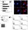
(A) Equivalent numbers (15,000) of vector-control and +HOXA10 T29-GFP cells were seeded onto wells coated with the indicated ECM proteins. At 1 h thereafter, attached T29-GFP cells counted in three random fields of 0.2 mm2 in each of three replicate wells. (B) Immunofluorescence staining of αvβ3 and α2β1 integrins. Bar, 20 μm. (C) Equivalent numbers (15,000) of vector-control and +HOXA10 T29-GFP cells were pre-incubated with no Ab, mouse IgG, αvβ3 Ab, and α2β1 Ab, and seeded onto confluent monolayers of omental mesothelial cells. At 2 h thereafter, numbers of attached T29-GFP cells were counted as described in (A). Adhesion assays were performed in triplicate in two independent experiments. (D) Detection of transcripts of β3 and αv integrins in T29 clones by semi-quantitative RT-PCR. (E) Flow cytometric analysis of staining of β3, αv and β1 integrins (shaded regions), and negative control staining with secondary Ab alone (dotted lines) in T29 cells.
Fig.4
Fig.4. HOXA10 promotes interactions of OSE cells with omental fibroblasts
(A) Equivalent numbers (15,000) of vector-control and +HOXA10 T29-GFP cells were seeded onto confluent monolayers of omental fibroblasts. At 2 h thereafter, attached T29-GFP cells were counted in three random fields of 0.2 mm2 in each of three replicate wells. (B) Adhesion assays were likewise performed where T29-GFP cells were pre-incubated with no Ab, mouse IgG, αvβ3 Ab, and α2β1 Ab, and then seeded onto fibroblast monolayers. In (C-E), cells of each T29-GFP line (1,000) were seeded with 1,000 omental fibroblasts. After direct co-culture for the indicated times, cells were stained with DAPI and viewed by immunofluorescence microscopy. Shown in (C) are co-cultures of T29-GFP cells (GFP+, DAPI+) and fibroblasts (GFP−, DAPI+) on Day 5. Bar, 50 μm. Total numbers of (D) T29-GFP cells and (E) fibroblasts in each well were calculated based on cell counts in three random fields of 0.2 mm2 in each of three replicate wells. In (F), 2,000 fibroblasts and 2,000 T29-GFP cells were seeded in the upper and lower chambers, respectively, of transwell co-culture plates. After indirect co-culture for the indicated times, the total number of T29-GFP cells in each well was determined by direct cell counts. All assays were performed in triplicate in two independent experiments.

References

    1. Anderson AR, Weaver AM, Cummings PT, Quaranta V. Tumor morphology and phenotypic evolution driven by selective pressure from the microenvironment. Cell. 2006;127:905–915. - PubMed
    1. Auersperg N, Wong AS, Choi KC, Kang SK, Leung PC. Ovarian surface epithelium: biology, endocrinology, and pathology. Endocr. Rev. 2001;22:255–288. - "VSports最新版本" PubMed
    1. Bei L, Lu Y, Bellis SL, Zhou W, Horvath E, Eklund EA. Identification of a HoxA10 activation domain necessary for transcription of the gene encoding beta3 integrin during myeloid differentiation. J. Biol. Chem. 2007;282:16846–16859. - PubMed
    1. Benson GV, Lim H, Paria BC, Satokata I, Dey SK, Maas RL. Mechanisms of reduced fertility in Hoxa-10 mutant mice: uterine homeosis and loss of maternal Hoxa-10 expression. Development. 1996;122:2687–2696. - PubMed (V体育官网入口)
    1. Carreiras F, Denoux Y, Staedel C, Lehmann M, Sichel F, Gauduchon P. Expression and localization of alpha v integrins and their ligand vitronectin in normal ovarian epithelium and in ovarian carcinoma. Gynecol. Oncol. 1996;62:260–267. - PubMed

Publication types

MeSH terms (VSports手机版)

V体育安卓版 - Grants and funding