Skip to main page content
U.S. flag

An official website of the United States government

Dot gov

The . gov means it’s official. Federal government websites often end in . gov or . mil. Before sharing sensitive information, make sure you’re on a federal government site. VSports app下载.

Https

The site is secure. The https:// ensures that you are connecting to the official website and that any information you provide is encrypted and transmitted securely V体育官网. .

. 2010 Feb;59(2):347-57.
doi: 10.2337/db09-0016. Epub 2009 Nov 23.

"V体育2025版" Depletion of liver Kupffer cells prevents the development of diet-induced hepatic steatosis and insulin resistance

Affiliations

"VSports最新版本" Depletion of liver Kupffer cells prevents the development of diet-induced hepatic steatosis and insulin resistance

Wan Huang et al. Diabetes. 2010 Feb.

Abstract

Objective: Increased activity of the innate immune system has been implicated in the pathogenesis of the dyslipidemia and insulin resistance associated with obesity and type 2 diabetes. In this study, we addressed the potential role of Kupffer cells (liver-specific macrophages, KCs) in these metabolic abnormalities VSports手机版. .

Research design and methods: Rats were depleted of KCs by administration of gadolinium chloride, after which all animals were exposed to a 2-week high-fat or high-sucrose diet. Subsequently, the effects of these interventions on the development of hepatic insulin resistance and steatosis were assessed V体育安卓版. In further studies, the effects of M1-polarized KCs on hepatocyte lipid metabolism and insulin sensitivity were addressed. .

Results: As expected, a high-fat or high-sucrose diet induced steatosis and hepatic insulin resistance V体育ios版. However, these metabolic abnormalities were prevented when liver was depleted of KCs. In vitro, KCs recapitulated the in vivo effects of diet by increasing hepatocyte triglyceride accumulation and fatty acid esterification, and decreasing fatty acid oxidation and insulin responsiveness. To address the mechanisms(s) of KC action, we inhibited a panel of cytokines using neutralizing antibodies. Only neutralizing antibodies against tumor necrosis factor-alpha (TNFalpha) attenuated KC-induced alterations in hepatocyte fatty acid oxidation, triglyceride accumulation, and insulin responsiveness. Importantly, KC TNFalpha levels were increased by diet in vivo and in isolated M1-polarized KCs in vitro. .

Conclusions: These data demonstrate a role for liver macrophages in diet-induced alterations in hepatic lipid metabolism and insulin sensitivity, and suggest a role for these cells in the etiology of the metabolic abnormalities of obesity/type 2 diabetes VSports最新版本. .


Figures

FIG. 1.
FIG. 1.
The effects of KC depletion on liver triglycerides, DAG, ceramide, and cholesterol in response to a high-fat or high-sucrose diet. Male Wistar rats depleted of KCs or control rats were exposed to a high-fat or high-sucrose diet for 2 weeks as described in research design and methods. Subsequently, animals were killed after an overnight fast, livers were isolated, and triglyceride (A), DAG (B), cholesterol (C), and ceramide (D) content of the liver was determined. Data are presented as means ± SE. Statistical significance is indicated. n = a minimum of six in each group.
FIG. 2.
FIG. 2.
The effects of KC depletion on the development of liver insulin resistance on a high-fat or high-sucrose diet. Male Wistar rats depleted of KCs or control rats were exposed to a high-fat (A and B) or high-sucrose (C and D) diet for 2 weeks. Subsequently, animals were fasted overnight and then underwent 4 mU · kg−1 · min−1 euglycemic-hyperinsulinemic clamp in the presence of [3-H3]-glucose for the measurement of hepatic insulin sensitivity as described in research design and methods. Hepatic glucose output (A and C) and insulin suppression of hepatic glucose output (B and D) were assessed. Data are presented as means ± SE. Statistical significance is indicated. n = a minimum of four in each group.
FIG. 3.
FIG. 3.
KC depletion by GdCl3 in liver and adipose tissue. Male Wistar rats were depleted of KCs by administration of GdCl3 as described in research design and methods. Subsequently, liver and adipose tissue were isolated and the presence of macrophages was determined by qRT-PCR assessment of CD68/ED1 and F4/80 mRNA levels (A and B) and counting of CD68/ED1-positive cells after immunohistochemical staining of liver and adipose tissue sections (C–F) as described in research design and methods. Statistical significance is indicated. n = a minimum of six in each group for qRT-PCR and n = 3 for immunohistochemical analysis of CD68/ED1-positive cells. (A high-quality digital representation of this figure is available in the online issue.)
FIG. 4.
FIG. 4.
Expression of lipid metabolism genes in the liver of SC-, HF-, and HS-fed rats with or without GdCl3 administration. Male Wistar rats were depleted of KCs by administration of GdCl3 as described in research design and methods. Subsequently, liver was isolated, RNA was extracted, and the expression of a number of lipid metabolism genes was determined by qRT-PCR. Statistical significance is indicated. n = a minimum of four in each group.
FIG. 5.
FIG. 5.
The effects of M1-polarized KCs on hepatocyte fatty acid oxidation and esterification and triglyceride accumulation. Hepatocytes and KCs were isolated from rat livers, plated, and cultured as described in research design and methods. Subsequently, the effects of the indicated exposures on fatty acid oxidation, fatty acid esterification, and triglyceride levels (A–C) and ACC and AMPK phosphorylation (D–F) were assessed as described in research design and methods. Data are presented as means ± SE. Statistical significance is indicated. n = a minimum of six in triplicate for each metabolic measurement. n = a minimum of four for ACC and AMPK analysis.
FIG. 6.
FIG. 6.
The effects of M1-polarized KCs on hepatocyte insulin responsiveness. Hepatocytes and KCs were isolated from rat livers, plated, and cultured as described in research design and methods. After the indicated exposures, hepatocytes were incubated with 100 nmol/l insulin for 10 min, and protein extracts were prepared and insulin receptor (IR) phosphorylation, IRS-1 phosphorylation, IRS-1–associated PI 3-kinase activity, and Akt phosphorylation were assessed as described in research design and methods. Data are presented as means ± SE. Statistical significance is indicated. n = a minimum of four for each experimental condition.
FIG. 7.
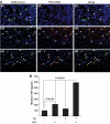
FIG. 7.
TNFα expression in KCs in liver in response to standard chow, high-fat, and high-sucrose diets and TNFα secretion from isolated M1-polarized KCs. Male Wistar rats were exposed to a high-fat or high-sucrose diet for 2 weeks. Subsequently, the livers were isolated, sections were prepared, and immunofluorescent staining for KCs and TNFα was performed (A), as described in research design and methods. The left panel shows CD68/ED1-positive cells (green), the center panels show TNFα-positive cells (red), and the right panels show the merged images. In all panels, Hoechst staining was used to visualize cell nuclei (blue). n = a minimum of three. For in vitro experiments (B), hepatocytes and KCs were isolated from rat livers, plated, and cultured as shown. After the indicated exposures, media were taken and TNFα was quantified by enzyme-linked immunosorbent assay (ELISA). Data are presented as means ± SE. Statistical significance is indicated. n = a minimum of three in each group. (A high-quality digital representation of this figure is available in the online issue.)
FIG. 8.
FIG. 8.
The effects of TNFα depletion on hepatocyte lipid metabolism and insulin-stimulated PI 3-kinase activity. Hepatocytes and KCs were isolated from rat livers, plated, and cultured as described in research design and methods. Subsequently, the effects of the indicated exposures on fatty acid oxidation and triglycerides in the absence or presence of a TNFα-neutralizing antibody were assessed as described in research design and methods (A). In separate experiments, hepatocytes were incubated with 100 nmol/l insulin for 10 min after similar exposures to those in (A), proteins extracts were then prepared, and IRS-1–associated PI 3-kinase activity was assessed as described in research design and methods (B). Data are presented as means ± SE. Statistical significance is indicated. n = a minimum of six for each experimental condition, except for triglycerides (n = 3).

References

    1. Hotamisligil GS, Erbay E: Nutrient sensing and inflammation in metabolic diseases. Nat Rev Immunol 2008;8:923–934 - PMC - PubMed
    1. Schenk S, Saberi M, Olefsky JM: Insulin sensitivity: modulation by nutrients and inflammation. J Clin Invest 2008;118:2992–3002 - PMC - PubMed
    1. Shoelson SE: Banking on ATM as a new target in metabolic syndrome. Cell Metab 2006;4:337–338 - PubMed
    1. Shoelson SE, Lee J, Goldfine AB: Inflammation and insulin resistance. J Clin Invest 2006;116:1793–1801 - V体育2025版 - PMC - PubMed
    1. Medzhitov R: Origin and physiological roles of inflammation. Nature 2008;454:428–435 - PubMed

VSports app下载 - Publication types

"V体育安卓版" MeSH terms