Skip to main page content
U.S. flag

An official website of the United States government

Dot gov

The . gov means it’s official VSports app下载. Federal government websites often end in . gov or . mil. Before sharing sensitive information, make sure you’re on a federal government site. .

Https

The site is secure. The https:// ensures that you are connecting to the official website and that any information you provide is encrypted and transmitted securely. V体育官网.

. 2009 Nov 24;10(1):118.
doi: 10.1186/1465-9921-10-118.

Characterisation of the proximal airway squamous metaplasia induced by chronic tobacco smoke exposure in spontaneously hypertensive rats

Affiliations

"VSports在线直播" Characterisation of the proximal airway squamous metaplasia induced by chronic tobacco smoke exposure in spontaneously hypertensive rats

Sarah J Bolton et al. Respir Res. .

Abstract

Background: Continuous exposure to tobacco smoke (TS) is a key cause of chronic obstructive pulmonary disease (COPD), a complex multifactorial disease that is difficult to model in rodents. The spontaneously hypertensive (SH) rat exhibits several COPD-associated co-morbidities such as hypertension and increased coagulation. We have investigated whether SH rats are a more appropriate animal paradigm of COPD. VSports手机版.

Methods: SH rats were exposed to TS for 6 hours/day, 3 days/week for 14 weeks, and the lung tissues examined by immunohistochemistry V体育安卓版. .

Results: TS induced a CK13-positive squamous metaplasia in proximal airways, which also stained for Ki67 and p63. We hypothesise that this lesion arises by basal cell proliferation, which differentiates to a squamous cell phenotype. Differences in staining profiles for the functional markers CC10 and surfactant D, but not phospho-p38, indicated loss of ability to function appropriately as secretory cells. Within the parenchyma, there were also differences in the staining profiles for CC10 and surfactant D, indicating a possible attempt to compensate for losses in proximal airways. In human COPD sections, areas of CK13-positive squamous metaplasia showed sporadic p63 staining, suggesting that unlike the rat, this is not a basal cell-driven lesion. V体育ios版.

Conclusion: This study demonstrates that although proximal airway metaplasia in rat and human are both CK13+ and therefore squamous, they potentially arise by different mechanisms VSports最新版本. .


Figures
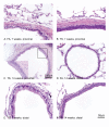
Figure 1
Figure 1
Morphology of TS induced changes in proximal and distal airways of SH rats. H&E stain for pathological assessment. Morphological changes were seen in proximal (A-C) and distal airways (D-F) following TS exposure. FA exposed rats showed minimal changes to proximal (A) and distal (D) airways. Exposure to TS for 7 (B) and 14 (C) weeks induced a squamous metaplasia in proximal airways (see high power inset in panel C). Distal airways showed epithelial hypertrophy at 7 weeks (E) but progressed to squamous metaplasia in occasional airways by 14 weeks (F). Magnification bar in F applies to panels A, B, D-F.
Figure 2
Figure 2
Morphology of TS induced changes in the lung parenchyma of SH rats. H&E stain for pathological assessment. FA exposed rats showed fibrinoid leakage from the alveolar bed (A, arrows) but minimal changes to resident cells. Following TS exposure for 14 week, there was evidence of an inflammatory infiltrate within perivascular/peribronchiolar regions (B, arrow). There was also fibrosis of blood vessels and the alveolar bed (C, arrows). Casts of the alveolar capillaries (C, inset, arrows) indicated remodelling of the microvascular network. Loss of connectivity was also seen (D, asterisks) within the alveolar bed.
Figure 3
Figure 3
Cellular turnover following TS exposure in SH rats. Antibodies to Ki67 (A-D) and cleaved caspase 3 (E, F) were used to determine the level of cell turnover following TS exposure. Areas of squamous metaplasia in both proximal (A) and distal (B) airways were strongly and extensively stained for Ki67 compared to FA controls (data not shown). Within the alveolar bed, the numbers of cells staining for Ki67 was increased after TS exposure (C) compared to the FA controls (D). Staining for cleaved capsase 3 showed a marginal increase in the numbers of cells staining following TS exposure (E, arrowheads) compared to FA controls (F, arrowhead). Staining was also seen in vesicles of alveolar macrophages (F, arrows). Magnification bar in B applies to panels A and B. Magnification bar in E applies to panels C-E.
Figure 4
Figure 4
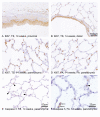
Epithelial profile of TS induced squamous metaplasia in proximal airways. Antibodies against pan CK (A, B), CK13 (C, D) and p63 (E, F, G) were used to characterise the squamous metaplastia seen in proximal airways following TS exposure. All epithelial cells were positive for panCK (A, B). After 7 weeks TS exposure, there was sporadic staining in areas of squamous metaplasia (C) but after 14 weeks, TS exposed rats showed CK13-positive staining in suprabasal regions of the entire airway (D). Staining for p63 highlighted sporadic basal stem cells in FA exposed rats (E, arrow). After 14 weeks of TS exposure in larger airways, all cell nuclei in areas of squamous metaplasia (F) were stained but with a gradation of intensity from the basal cells (intense) to the suprabasal cells (weak). Staining with the isotype control antibody (G) showed no specific nuclear staining although there was weak background staining in the cytoplasm.
Figure 5
Figure 5
Molecular profile of functional markers in the lung parenchyma of TS induced SH rats. CC10 (A, B), p-p38 (C, D) and SP-D (E, F) expression were examined in the lung parenchyma after FA (A, C, E) and TS (B, D, F) exposure. After FA exposure, CC10 was only seen in the parenchyma in occasional alveolar macrophages and type II pneumocytes. The image shown in A includes a transitional airway to demonstrate normal Clara cell staining. After TS exposure, CC10 was now seen more diffusely in the alveolar bed in type II cells and alveolar macrophages (B). p-p38 (C) and SP-D (E) were observed in macrophages and type II pneumocytes within the alveolar bed in FA exposed SH rats and staining pattern appeared more extensive after TS exposure and included type I pneumocytes (D, F).
Figure 6
Figure 6
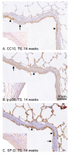
Association of functional markers with squamous metaplasia in distal airways. Antibodies against CC10 (A), p-p38 (B) and SP-D (C) were used to assess the effect of squamous metaplasia on epithelial cell functional markers. Within distal airways, there were areas of squamous metaplasia (arrow) contiguous with areas of non-squamous epithelial cells (arrowhead). CC10 (A) was lost from areas of squamous metaplasia (A, arrow), including the proximal airways (inset) but not from other areas of non-squamous airways (A, arrowhead). p-p38 (B, distal airway) was detected in all epithelial cells regardless of cell type including squamous metaplasia in the proximal airways (inset). SP-D (C) was lost from squamous regions in distal (C, arrow) and proximal airways (C, inset). In areas of non-squamous epithelium in the distal airways, epithelial cells were seen to stain positive (C, arrowhead).
Figure 7
Figure 7
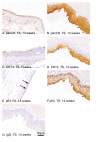
Comparison of squamous metaplasia in human COPD and TS exposed SH rat lungs. Squamous metaplasia was identified in human COPD lung tissue (C, E) and 14 week TS exposed SH rat lungs (D, F) and compared to either non-squamous airways in COPD lung (A) or FA exposed SH rats (B). They were stained for p63 (A, B, E, F) and CK13 (C, D). In the control non-squamous airways from both human (A) and rat (B), p63 stained occasional flattened cells close to the basement matrix of proximal airways. Areas of squamous metaplasia were identified in both human (C) and rat (D) tissue by CK13 staining. In contrast to the extensive p63 staining seen in the rat tissue (F), the human tissue showed only sporadic cells scattered through out the squamous metaplasia (E).

References

    1. Hogg JC. Pathophysiology of airflow limitation in chronic obstructive pulmonary disease. The Lancet. 2004;364:709–721. doi: 10.1016/S0140-6736(04)16900-6. - DOI - PubMed
    1. Kim V, Rogers TJ, Criner GJ. New Concepts in the Pathobiology of Chronic Obstructive Pulmonary Disease. Proc Am Thorac Soc. 2008;5:478–485. doi: 10.1513/pats.200802-014ET. - "VSports" DOI - PMC - PubMed
    1. Lapperre T, Sont J, van Schadewijk A, Gosman M, Postma D, Bajema I, Timens W, Mauad T, Hiemstra P. the GLUCOLD Study Group. Smoking cessation and bronchial epithelial remodelling in COPD: a cross-sectional study. Resp Res. 2007;8:85. doi: 10.1186/1465-9921-8-85. - DOI - PMC - PubMed
    1. Puchelle E, Zahm JM, Tournier JM, Coraux C. Airway Epithelial Repair, Regeneration, and Remodeling after Injury in Chronic Obstructive Pulmonary Disease. Proc Am Thorac Soc. 2006;3:726–733. doi: 10.1513/pats.200605-126SF. - DOI - PubMed
    1. Rennard SI. Inflammation and Repair Processes in Chronic Obstructive Pulmonary Disease. Am J Respir Crit Care Med. 1999;160:12S–16. - "V体育安卓版" PubMed

Publication types

"V体育官网入口" MeSH terms