Skip to main page content
U.S. flag

An official website of the United States government

Dot gov

The . gov means it’s official. Federal government websites often end in . gov or VSports app下载. mil. Before sharing sensitive information, make sure you’re on a federal government site. .

Https

The site is secure. The https:// ensures that you are connecting to the official website and that any information you provide is encrypted and transmitted securely V体育官网. .

. 2009 Dec 1;183(11):6939-47.
doi: 10.4049/jimmunol.0902000. Epub 2009 Nov 13.

Chimeric NKG2D expressing T cells eliminate immunosuppression and activate immunity within the ovarian tumor microenvironment

Affiliations

Chimeric NKG2D expressing T cells eliminate immunosuppression and activate immunity within the ovarian tumor microenvironment

Amorette Barber et al. J Immunol. .

V体育平台登录 - Erratum in

  • J Immunol. 2014 Aug 1;193(3):1513

Abstract

Adoptive transfer of T cells expressing chimeric NKG2D (chNKG2D) receptors, a fusion of NKG2D and CD3zeta, can lead to long-term, tumor-free survival in a murine model of ovarian cancer. To determine the mechanisms of chNKG2D T cell antitumor efficacy, we analyzed how chNKG2D T cells altered the tumor microenvironment, including the tumor-infiltrating leukocyte populations. chNKG2D T cell treatment of mice bearing ID8 tumor cells increased the number and activation of NK cells and increased the activation of host CD8+ T cells within the tumor. Foxp3+ regulatory T cells at the tumor site decreased more than 300-fold after chNKG2D T cell treatment. Tumor-associated regulatory T cells expressed cell surface NKG2D ligands and were killed by chNKG2D T cells in a perforin-dependent manner. chNKG2D T cells also altered the function of myeloid cells at the tumor site, changing these cells from being immunosuppressive to enhancing T cell responses. Cells isolated from the tumor produced elevated amounts of IFN-gamma, NO, and other proinflammatory cytokines after chNKG2D T cell treatment. ChNKG2D T cells required perforin, IFN-gamma, and GM-CSF to induce a full response at the tumor site. In addition, transfer of chNKG2D T cells into mice bearing tumors that were established for 5 weeks led to long-term survival of the mice. Thus, chNKG2D T cells altered the ovarian tumor microenvironment to eliminate immunosuppressive cells and induce infiltration and activation of antitumor immune cells and production of inflammatory cytokines VSports手机版. This induction of an immune response likely contributes to chNKG2D T cells' ability to eliminate established tumors. .


Figures
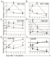
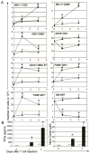
Figure 1
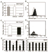
Figure 1. Treatment with chNKG2D T cells induced activation of the tumor-infiltrating leukocyte populations
ID8-GFP cells were injected and mice were treated with Ly5.1+ wtNKG2D (triangles) or chNKG2D (circles) T cells i.p. seven days later. (A) The number of NK1.1+CD3 NK cells (B) CD69+NK1.1+CD3 activated NK cells, (C) Ly5.1 CD8+ CD69+ T cells, (D) F480 GR1+ neutrophils, (E) CD19+ CD3 B cells, or (F) GFP+ ID8 tumor cells was determined prior to T cell injection (day 0), and one, three, and seven days after T cell injection. The average of each group (n=4) is shown. Treatment with chNKG2D T cells significantly changed the number of the different cell populations compared to control treated mice (*-p<0.05). Data are representative of at least 5 independent experiments.
Figure 2
Figure 2. ChNKG2D T cell treatment decreases Foxp3+ CD4 T cells at the tumor site
ID8-GFP cells were injected i.p. into mice. After eight weeks mice received no treatment (-, grey bars), or were treated with Ly5.1+ wtNKG2D (white bars) or chNKG2D (black bars) T cells i.p. Three days after wtNKG2D or chNKG2D T cell injection, the number of Ly5.1 Foxp3+CD4+ T cells was determined in (A) the peritoneal wash or (C) the spleen. The average of each group + SD (n=4) is shown. Treatment with chNKG2D T cells significantly decreased the number of the Foxp3+CD4+ T cells in the peritoneal cavity compared to control treated mice (***- p<0.001). (B and D) The expression of NKG2D ligands on Foxp3+CD4+ T cells was determined by staining (B) peritoneal wash cells or (D) spleen cells with sNKG2D-hIgG (filled) or human IgG isotype control (grey) and with anti-human IgG secondary antibodies. The FACS histograms show NKG2D ligand expression on Ly5.1 CD3+CD4+Foxp3+ cells and are representative of three separate experiments, with n=4 for each experiment. (E and F) ID8-GFP cells were injected i.p. into mice. After eight weeks, peritoneal wash cells were cultured with media, wtNKG2D T cells, chNKG2D T cells, or chNKG2D T cells deficient in perforin (Pfp), FasL, or perforin and FasL (P/F). After 24 hours, the percent of (E) Foxp3+CD4+ T cells or (F) CD19+CD3 cells was determined. Data are representative of two independent experiments. Culture with chNKG2D T cells significantly decreased the percent of Foxp3+CD4+ T cells (*- p<0.001).
Figure 3
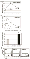
Figure 3. Antigen presenting cells at the tumor site are activated after chNKG2D T cell injection
ID8-GFP cells were injected i.p. into mice. After seven days mice were treated with wtNKG2D (triangles) or chNKG2D (circles) T cells i.p. (A) The number of CD11c+MHC class IIhi cells or (B) the number of F4/80+GR1+ cells was determined prior to T cell injection (day 0), and one, three, and seven days after T cell injection. (C-D) F4/80+ cells were isolated from the peritoneal washes three days after injection of wtNKG2D (white bars) or chNKG2D (black bars) T cells or from naïve mice (grey bars) and were cultured with CFSE-labeled OT-I T cells and Ova peptide. OT-I T cell proliferation was measured after four days of culture. (C) The average of each group + SD (n=4) and (D representative histograms of OT-I T cell proliferation are shown. Treatment with chNKG2D T cells significantly changed the number of the different cell populations compared to control treated mice (*-p<0.05). Data are representative of at least 2 separate experiments.
Figure 4
Figure 4. Tumor infiltrating cells from chNKG2D T cell treated mice have increased IFNγ and nitric oxide secretion
(A) Peritoneal wash cells from wtNKG2D (white bars) or chNKG2D T cell (black bars) treated tumor-bearing mice from each timepoint were cultured in media for 24 hours. Cell-free supernatants were assayed for (A) IFNγ or nitric oxide. (B) Intracellular staining was performed on peritoneal wash cells cultured in media for 24 hours. Cells were evaluated for IFNγ production and were gated on either Ly5.1+CD3+, CD8+CD3+, CD4+CD3+, or NK1.1+CD3 as indicated. The average of each group + SD (n=4) is shown. Treatment with chNKG2D T cells significantly increased IFNγ and nitric oxide secretion compared to control treated mice (*- p<0.05, **-p<0.01). Data are representative of at least 5 separate experiments.
Figure 5
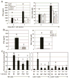
Figure 5. ChNKG2D T cell-derived GM-CSF, IFNγ, and perforin are required for the increase in IFNγ and nitric oxide production in tumor-bearing mice
(A) Peritoneal wash cells from B6- derived wtNKG2D (white bars) or chNKG2D T cells (black bars), or chNKG2D T cells deficient in GM-CSF (hashed), IFNγ (striped), or perforin (grey) treated tumor bearing mice from each timepoint were cultured in media for 24 hours. Cell-free supernatants were assayed for (A) IFNγ or nitric oxide. (B) Three days after T cell injection, peritoneal wash cells from B6 mice treated with wtNKG2D (white bars) or chNKG2D T cells (black bars), or IFNγR−/− mice treated with wtNKG2D (hashed bars) or chNKG2D T cells (grey) were cultured in media for 24 hours. Cell-free supernatants were assayed for IFNγ or nitric oxide. (C) Intracellular staining was performed on peritoneal wash cells cultured in media for 24 hours. Cells were evaluated for IFNγ production and were gated on either CD8+CD3+, CD4+CD3+, or NK1.1+CD3 as indicated. The average of each group + SD (n=4) is shown. Treatment with chNKG2D T cells significantly increased IFNγ and nitric oxide secretion compared to control treated mice (*p<0.05) and mice treated with chNKG2D T cells deficient in effector molecules produced significantly less IFNγ and nitric oxide compared to chNKG2D T cell treated mice (§- p<0.05). Data are representative of at least 2 separate experiments.
Figure 6
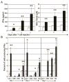
Figure 6. Treatment of advanced tumors with chNKG2D T cells induced activation of the tumor-infiltrating leukocyte populations
ID8-GFP cells were injected, and mice were treated with wtNKG2D (triangles) or chNKG2D (circles) T cells i.p. five weeks later. (A) The number of NK1.1+CD3 NK cells, CD69+NK1.1+CD3 activated NK cells, Ly5.1CD8+ CD69+ T cells, CD19+ CD3 B cells, CD11c+MHC class IIhi cells, F4/80+GR1+ cells, F480 GR1+ neutrophils,or GFP+ ID8 tumor cells was determined prior to T cell injection (day 0), and one and three days after T cell injection. (B) Peritoneal wash cells from wtNKG2D (white bars) or chNKG2D T cell (black bars) treated tumor-bearing mice from each timepoint were cultured in media for 24 hours. Cell-free supernatants were assayed for IFNγ or nitric oxide. The average of each group (n=4) is shown. Treatment with chNKG2D T cells significantly changed the number of the different cell populations compared to control treated mice (*-p<0.05). Data are representative of 2 independent experiments.
Figure 7
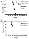
Figure 7. ChNKG2D T cells increase survival of mice bearing established ID8 tumors
ID8-GFP cells were injected on day 0, and mice were treated three times with wtNKG2D (filled symbols) or chNKG2D (open symbols) T cells i.p. either (A) after five, six, and seven weeks or (B) after five, seven, and nine weeks. The survival of the mice was measured (n=11–12 per group). Treatment with chNKG2D T cells significantly increased the survival of tumor-bearing mice compared to control treated mice (***- p<0.001).

References

    1. Rosenberg SA, Restifo NP, Yang JC, Morgan RA, Dudley ME. Adoptive cell transfer: a clinical path to effective cancer immunotherapy. Nat Rev Cancer. 2008;8:299–308. - PMC - PubMed
    1. Cohen JI, Bollard CM, Khanna R, Pittaluga S. Current understanding of the role of Epstein-Barr virus in lymphomagenesis and therapeutic approaches to EBV-associated lymphomas. Leuk Lymphoma. 2008;49(Suppl 1):27–34. - PMC - PubMed
    1. June CH. Adoptive T cell therapy for cancer in the clinic. J Clin Invest. 2007;117:1466–1476. - PMC - PubMed
    1. Kershaw MH, Westwood JA, Parker LL, Wang G, Eshhar Z, Mavroukakis SA, White DE, Wunderlich JR, Canevari S, Rogers-Freezer L, Chen CC, Yang JC, Rosenberg SA, Hwu P. A phase I study on adoptive immunotherapy using gene-modified T cells for ovarian cancer. Clin Cancer Res. 2006;12:6106–6115. - "VSports app下载" PMC - PubMed
    1. Nelson BH. The impact of T-cell immunity on ovarian cancer outcomes. Immunol Rev. 2008;222:101–116. - PubMed

Publication types

MeSH terms

Substances