Skip to main page content
U.S. flag

An official website of the United States government

Dot gov

The VSports app下载. gov means it’s official. Federal government websites often end in . gov or . mil. Before sharing sensitive information, make sure you’re on a federal government site. .

Https

The site is secure. The https:// ensures that you are connecting to the official website and that any information you provide is encrypted and transmitted securely V体育官网. .

. 2009 Dec;15(12):1513-22.
doi: 10.1016/j.bbmt.2009.08.013. Epub 2009 Oct 7.

"VSports手机版" Blocking LFA-1 activation with lovastatin prevents graft-versus-host disease in mouse bone marrow transplantation

Affiliations

"V体育2025版" Blocking LFA-1 activation with lovastatin prevents graft-versus-host disease in mouse bone marrow transplantation

VSports在线直播 - Yang Wang et al. Biol Blood Marrow Transplant. 2009 Dec.

Abstract

Graft-versus-host disease (GVHD) following bone marrow transplantation (BMT) is mediated by alloreactive donor T lymphocytes VSports手机版. Migration and activation of donor-derived T lymphocytes play critical roles in the development of GVHD. Leukocyte function-associated antigen-1 (LFA-1) regulates T cell adhesion and activation. We previously demonstrated that the I-domain, the ligand-binding site of LFA-1, changes from the low-affinity state to the high-affinity state on LFA-1 activation. Therapeutic antagonists, such as statins, inhibit LFA-1 activation and immune responses by modulating the affinity state of the LFA-1 I-domain. In the present study, we report that lovastatin blocked mouse T cell adhesion, proliferation, and cytokine production in vitro. Furthermore, blocking LFA-1 in the low-affinity state with lovastatin reduced the mortality and morbidity associated with GVHD in a murine BMT model. Specifically, lovastatin prevented T lymphocytes from homing to lymph nodes and Peyer's patches during the GVHD initiation phase and after donor lymphocyte infusion (DLI) after the establishment of GVHD. In addition, treatment with lovastatin impaired donor-derived T cell proliferation in vivo. Taken together, these results indicate the important role of lovastatin in the treatment of GVHD. .


Figures
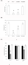
Figure 1
Figure 1. Lovastatin inhibits the adhesion, proliferation and cytokine production of mouse T cells in vitro
(A) Blocking of mouse T cell binding to ICAM-1 with lovastatin. Primary mouse T cells (WT or LFA-1−/−) were activated with Mn++, and WT were pre-incubated with lovastatin or pravastatin at the concentration of 10uM. Binding to ICAM-1 was measured by counting cells adherent to the wells after washes. (B and C) Inhibition of mouse T cell proliferation (B) and cytokine production (C) with lovastatin. Column purified C57BL/6 responder cells were plated at 1×10⁶ cells/ml in a volume of 200 μl/well and cocultured at a ratio of 2:1 with 3400 cGy irradiated stimulator cells from the Balb/C mice. Proliferation was assayed on day 3 by adding [3H]-thymidine to the culture for the last 8 hours. Production of IL-2, TNF-α and IFN-γ in culture supernatant were measured by ELISA after 24 hrs in the presence of DMSO (black bar), lovasatin (gray bar) and pravastatin (white bar). Results are mean and S.D. of three independent experiments normalized to that of WT control. Asterisk represents data with p value less than 0.05 in t test.
Figure 2
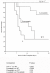
Figure 2. Lovastatin treatment reduces mortality associated with GVHD
Mice were treated with statins at the dose of 50 μg/mouse every other day, starting on the same day as the BMT. The control group received vehicle (10% DMSO in saline). Survival was monitored daily and followed up until 28 days. The data was the combination of all the mice examined. Distributions of time to death were estimated using the Kaplan-Meier method and compared between treatments by using the log-rank Test. No adjustment was made for the comparisons. Results were from at least three independent experiments with 3 mice per group in each experiment.
Figure 3
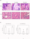
Figure 3. Lovastatin treatment Reduces GVHD in skin and liver
Mice were sacrificed on day 7 post-transplant (4 mice per group). Tissues were placed in 10% formalin, embedded in paraffin, sectioned, and stained with hematoxylin and eosin, and scored for GVHD histopathology. (A) The top panels are representative sections from the skin of mice treated with DMSO, lovastatin and pravastatin. Skin shows moderate to severe changes consistent with GVHD with lymphoid infiltrates (arrow), epidermal and adnexal cell apoptosis in WT and pravastatin-treated mice. There were only mild changes noted in the skin of lovastatin-treated mice. The lower panels are representative sections from the liver. Perivascular lymphoid infilrates (arrows) within portal triads associated with bile duct damage were noted in the liver of WT and pravastatin treated mice but not in the lovastatin-treated mice. (B) The average score of skin and liver GVHD of each group. Results are mean and S.D. of total 4 mice. Asterisk represents data with p value less than 0.05 in t test.
Figure 4
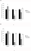
Figure 4. Donor-derived T cell homing to the secondary lymphoid organs
Donor-derived splenocytes were labeled with CFSE and transferred into mice on the same day of transplantation immediately after statin treatment. The recipient mice were sacrificed 2 hours after transfer. Cells were collected from spleen, peripheral lymph nodes and Peyer’s patches, then stained with CD4-PerCP, CD8-APC and H-2Db-PE antibodies. The number of injected T cell subsets recovered from each tissue, CD4+/H-2Db+/CFSEhi (A) and CD8+/H-2Db+/CFSEhi(B), were determined by FACS. Results were calculated as the mean and S.D. of at least 3 independent experiments normalized to that of WT control. Asterisks, data with p value less than 0.05 in t test.
Figure 5
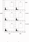
Figure 5. Donor-derived T cell proliferation in the spleen
The in vivo proliferation of donor-derived T cells in the spleen was measured by CFSE tracking assay. Donor splenocytes were labeled with CFSE and then infused into the recipients with the bone marrow cells as described. Mice were sacrificed on day 3 post-transplant. Cells from spleen were stained with CD4-PerCP, CD8-APC and H-2Db-PE. The CD4+ (left panel) or CD8+ (right panel) cells were displayed on dot plots for the donor marker H-2Db. The percentage of donor-derived cells (CD4+/H-2Db+ or CD8+/H-2Db+) was labeled on the top of the boxed region, and the lower-left insert of each plot represented the CFSE histogram of this designated population. Data shown here are representative of at least 3 independent experiments.
Figure 6
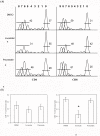
Figure 6. Donor-derived T cell proliferation in the peripheral lymph nodes
The in vivo proliferation of donor-derived T cells in the lymph nodes was measured on day 4 post-transplant using the CFSE tracking assay. Cells from lymph nodes were stained with CD4-PerCP, CD8-APC and H-2Db-PE. The donor-derived T lymphocyte subsets (CD4+/H-2Db+ and CD8+/H-2Db+) were determined by FACS. (A) The proliferation kinetics of donor-derived CD4+ (left panel) or CD8+ (right panel) cells were analyzed by FlowJo. The cell proliferation models were generated based on the CFSE histogram data. The cell division numbers were displayed on the top of each panel. The percentages of undivided cells and dividing cells were labeled in each figure. (B) Cell division index were calculated by FlowJo based on the proliferation kinetics. Data shown here are representative of 3 independent experiments. Asterisks, data with p value less than 0.05 in t test.

"V体育安卓版" References

    1. Sale GE, Shulman HM, Hackman RC. Pathology of hematopoietic cell transplantation. In: Blume KG, Forman SJ, Appelbaum FR, editors. Thomas’s Hematopoietic Cell Transplantation. Blackwell Scientific Publications; Boston, MA: 2004. pp. 286–299.
    1. Antin JH, Deeg HJ. Clinical spectrum of acute graft-vs.-host disease. In: Ferrara JLM, Cooke KR, Deeg HJ, editors. Graft-vs.-Host-Disease. Marcel Dekker; New York, NY: 2005. pp. 369–382.
    1. Ferrara JLM, Cooke KR, Teshima T. The Pathophysiology of graft-vs.-host disease. In: Ferrara JLM, Cooke KR, Deeg HJ, editors. Graft-vs.-Host-Disease. Marcel Dekker; New York, NY: 2005. pp. 1–34.
    1. Korngold R, Friedman TM. Murine models for graft-versus-host disease. In: Blume KG, Forman SJ, Appelbaum FR, editors. Thomas’s Hematopoietic Cell Transplantation. Blackwell Scientific Publications; Boston, MA: 2004. pp. 353–368.
    1. Blazar BR, Korngold R, Vallera DA. Recent advances in graft-versus-host disease (GVHD) prevention. Immunol Rev. 1997;57:79–109. - "V体育官网" PubMed

MeSH terms (V体育2025版)