<b dir="fbFxYz"></b> "V体育官网入口" Skip to main page content
U.S. flag

An official website of the United States government

Dot gov

The . gov means it’s official. Federal government websites often end in VSports app下载. gov or . mil. Before sharing sensitive information, make sure you’re on a federal government site. .

Https

The site is secure. The https:// ensures that you are connecting to the official website and that any information you provide is encrypted and transmitted securely. V体育官网.

. 2010 Jan;24(1):177-86.
doi: 10.1038/leu.2009.224. Epub 2009 Nov 5.

WAVE1 regulates Bcl-2 localization and phosphorylation in leukemia cells

Affiliations

WAVE1 regulates Bcl-2 localization and phosphorylation in leukemia cells

R Kang et al. Leukemia. 2010 Jan.

Abstract

Bcl-2 proteins are over-expressed in many tumors and are critically important for cell survival. Their anti-apoptotic activities are determined by intracellular localization and post-translational modifications (such as phosphorylation). Here, we showed that WAVE1, a member of the Wiskott-Aldrich syndrome protein family, was over-expressed in blood cancer cell lines, and functioned as a negative regulator of apoptosis. Further enhanced expression of WAVE1 by gene transfection rendered leukemia cells more resistant to anti-cancer drug-induced apoptosis; whereas suppression of WAVE1 expression by RNA interference restored leukemia cells' sensitivity to anti-drug-induced apoptosis. WAVE1 was found to be associated with mitochondrial Bcl-2, and its depletion led to mitochondrial release of Bcl-2, and phosphorylation of ASK1/JNK and Bcl-2 VSports手机版. Furthermore, depletion of WAVE1 expression increased anti-cancer drug-induced production of reactive oxygen species in leukemia cells. Taken together, these results suggest WAVE1 as a novel regulator of apoptosis, and potential drug target for therapeutic intervention of leukemia. .


Figures

Figure 1
Figure 1. WAVE1 is over-expressed in human leukemia cell lines and renders cells resistant to chemotherapeutics- induced apoptosis
(A) WAVE1 was over-expressed in blood cancer cell lines. Western blotting analysis of WAVE1 and WAVE2, and GAPDH in various human cancer cell lines as indicated. (B) Relative expression levels of WAVE1 in childhood leukemia. Total protein was extracted from normal or patients' BMMCs, and equal amounts of proteins (30 μg) were loaded, and WAVE1 level was determined by the relative optical intensity (in arbitrary units, AU) of the immunoreactive bands on Western blots. Each dot represents relative WAVE1 level in each individual sample. ALL, acute lymphoblastic leukemia; ANLL, acute nonlymphocytic leukemia; P, primary; CR, complete remission; R, relapse. *P < 0.05 versus normal. #P < 0.05 versus CR. (C) WAVE1 knockdown increased the sensitivity of leukemia cells to anti-cancer drug induced apoptosis. Knockdown WAVE1 by siRNA in leukemia cells as indicated and then treated with VCR (1 μg/ml), ADM (1 μg/ml), and AS2O3 (5 μM). Cell apoptosis was examined at 24 and 48 h, and caspase-3 activity was examined at 24 h (n=3). UT, Untreated group. *P < 0.05 versus Control siRNA group. (D) WAVE1 overexpression rendered HL-60 leukemia cells resistant to anti-cancer drug -induced apoptosis. HL-60 cells transfected with WAVE1, WAVE2 and pEFBOS vector plasmid and then treat with VCR (1 μg/ml), ADM (1 μg/ml), and AS2O3 (5 μM). Cell apoptosis and caspase 3 activity was examined at 24 h (n=3). UT, Untreated group. *P < 0.05 versus empty vector group. (E) Knockdown of Bcl-2 by shRNA in WAVE1 over-expressing cells restored sensitivity to anti-cancer drug-induced apoptosis. HL-60 cells transfected with various plasmid were stimulated with VCR (1 μg/ml), ADM (1 μg/ml), and AS2O3 (5 μM). At 24 h post treatment, cell apoptosis was assayed (n=3, *, # P < 0.05).
Figure 2
Figure 2. WAVE1 regulates ROS production and Ca2+ homeostasis in apoptosis
(A) Knockdown WAVE1 by siRNA in Jurkat or HL-60 cells and treated with ADM (1 μg/ml), thapsigargin (100 nM),or H2O2 (250 μmol) for 24 h, and ROS production, ER Ca2+ release (cytosolic free calcium) and mitochondria Ca2+ load was analyzed by flow cytometry (n=3, * P < 0.05 versus control siRNA group). UT, Untreated group, set as 1. (B) Knockdown WAVE1 cells were treated with thapsigargin (100 nM) or H2O2 (250 μmol) with or without FCCP (2.5 μmol) for 24 h, and mitochondria Ca2+ load was analyzed by flow cytometry (n=3, * P < 0.05). Untreated group, set as 1. (C) Knockdown of Bcl-2 by shRNA in WAVE1 overexpressin cells restored ROS and Ca2+ production. HL-60 cells transfected with various plasmids were stimulated with thapsigargin (100 nM) for 24 h, and cellular levels of ROS, cytosolic free calcium, and mitochondria Ca2+ load was analyzed by flow cytometry (n = 3, * P < 0.05).
Figure 3
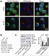
Figure 3. WAVE1 regulates intracellular localization of Bcl-2 in leukemia cell
(A) Localization of WAVE1 in leukemia cell. Jurkat and HL-60 cells were stained with WAVE1-specific antibodies (Green) and Mitotracker (Red) and nuclear Hoechst 33258 (Blue). (B) Localization of WAVE1 in mitochondria. Western blotting analysis of WAVE1 in cytoplasmic and mitochondrial fractions of Jurkat and HL-60 cells. The successful separation of cytoplasmic (“Cyt”) and mitochondria (“Mit”) fraction was confirmed by western blotting analysis of each fraction for known cytoplasmic (tubulin), mitochondria (mHSP70), nuclear (H3) and ER (PDI). (C) WAVE1 regulated Bcl-2 intracellular localization. Western blotting analysis of Bcl-2, Bcl-xL and Bad levels in cytoplasmic (“Cyt”) and mitochondria (“Mit”) fractions of Jurkat cells with or without WAVE siRNA or gene transfection. PDI and mHSP70 were used as fraction isolation quality control. * P < 0.05
Figure 4
Figure 4. WAVE1 binds Bcl-2 in mitochondria in leukemia cell
(A) CoIP assay Jurkat cells were transfected with plasmids encoding Myc-tagged WAVE1 and GFP-tagged Bcl-2, Bcl-xl, Bad and Bid. Cell lysates normalized for total protein content were analyzed directly or subjected to IP using anti-Myc or anti-GFP antibody and were analyzed by western blotting using anti-Myc or anti-GFP antibodies. (B) Endogenous WAVE1 bind Bcl-2. Jurkat cells were treat with or without ADM for 24 h, and lysates were prepared for IP with control IgG or anti-WAVE1, anti-Bcl-2, anti-Bcl-xl, anti-Bad, anti-Bid. The resulting immune complexes were analyzed by western blotting using antibodies recognizing WAVE1, Bcl-2, Bcl-xl, Bad, or Bid. (C) Jurkat cells were transfected with plasmids encoding GFP-tagged Bcl-2, mitochondria-restricted Bcl-2 (Mit-Bcl-2), ER-restricted Bcl-2 (ER-Bcl-2). Cells were fractionated into mitochondrial (Mit), and nuclear (ER), and were analyzed by western blotting. PDI and mHSP70 were used as fraction isolation quality control. (D) CoIP assay Jurkat cells were transfected with plasmids encoding Myc-tagged WAVE1 and GFP-tagged Bcl-2, mitochondria-restricted Bcl-2 (Mit-Bcl-2), and ER-restricted Bcl-2 (ER-Bcl-2). Cell lysates normalized for total protein content were analyzed directly or subjected to IP using anti-Myc or anti-GFP antibody and were analyzed by western blotting using anti-Myc or anti-GFP antibodies. (E) Endogenous WAVE1 bind mitochondria Bcl-2. The cytosolic and mitochondria fractions were prepared and lysates were prepared for IP with anti-Bcl-2. The resulting immune complexes were analyzed by western blotting using antibodies recognizing WAVE1. The inputs were analyzed by western blotting using anti-mHSP70 and tubulin.
Figure 5
Figure 5. WAVE1 regulates Bcl-2-mediated ROS production in leukemia cell
(A) Effects of WT Bcl-2, mitochondria-Bcl-2, and ER-Bcl-2 on ADM-induced cell death, ROS production, and caspase-3 activity with or without WAVE1 shRNA. Jukat cells were co-transfected with expression plasmids (Bcl-2-GFP, Mit-Bcl-2-GFP, and ER-Bcl-2-GFP) and/or shRNA plasmids (WAVE1 shRNA and control shRNA), then stimulated with ADM (1 μg/ml) for 24 h, and assayed ROS production, cell death and caspase-3 activity as indicated (n=3, * P < 0.05 versus vector group). (B) WAVE1 knockdown impaired mitochondria localization of nature Bcl-2, but not mitochondria-restricted Bcl-2. Jukat cells were co-transfected with expression plasmids (Bcl-2-GFP, Mit-Bcl-2-GFP, and ER-Bcl-2-GFP) and/or shRNA plasmids (WAVE1 shRNA and control shRNA), then isolated mitochondria, and western blotting assay GFP and mHSP70 levels. (C) No influence of WAVE1 on mitochondria localization of monoamine oxidase B (MAO-B) protein. Knockdown WAVE1 by shRNA in Jurkat cells, then isolated mitochondria, and western blotting assay MAO-B and mHSP70 levels.
Figure 6
Figure 6. WAVE1 regulates Bcl-2-mediated Ca2+ homeostasis in leukemia cell
Jurkat cells were co-transfected with expression plasmids (Bcl-2-GFP, Mit-Bcl-2-GFP, and ER-Bcl-2-GFP) and/or shRNA plasmids (WAVE1 shRNA and control shRNA), then stimulated with thapsigargin (100 nM) for 24 h, and assayed cell death, ER Ca2+ release (cytosolic free calcium) and mitochondria Ca2+ load, ROS production, pro-caspase-12 level, and caspase3 activity as indicated (n=3, * P < 0.05 versus vector group).
Figure 7
Figure 7. WAVE1 regulates Bcl-2 phosphorylation in an ASK1/JNK-dependent pathway
(A) WAVE1 knockdown increased Bcl-2 phosphorylation in ER. Knockdown WAVE1 by shRNA in Jurkat cells, then isolated mitochondria (Mit) and ER, and western blotting assay phosphorylation-Bcl-2 (p-Bcl-2) levels. The successful separation of cytoplasmic (“Cyt”) and mitochondria (“Mit”) fraction was confirmed by Western blotting analysis of each fraction for known mitochondrial (mHSP70, COXIV), nuclear (H3) and ER (PDI, Calnexin) markers. (B) Effects of Bcl-2 phosphorylation on ER stress-induced cell death. Jurkat cells expressing neo vector, Bcl-2 WT, or Bcl-2 AAA (non-phosphorylation Bcl-2) were treated thapsigargin (100 nM) for 24 h, and assayed cell death. * P < 0.05 versus Bcl-2 WT group. (C) Effects of Bcl-2 WT, Bcl-2 AAA with or without WAVE1 on ER stress-induced cell death. Jurkat cells were co-transfected with expression plasmids (Bcl-2-WT, and Bcl-2 AAA) and/or shRNA plasmids (WAVE1 shRNA and control shRNA), then stimulated with thapsigargin (100 nM) for 24 h, and assayed cell death. * P < 0.05 versus Bcl-2 WT group. (D) Effects of WAVE1 on ER stress-induced activation of JNK pathway. Jurkat cells expressing either control shRNA or WAVE1 shRNA vector were stimulated with thapsigargin at indicated dose for 30 min, and analyzed phosphorylation of JNK (p-JNK1/2), ASK1 (p-ASK1), p38 (p38), and RAC1 (p-RAC1) by western blotting. Data showed as the relative optical intensity (in arbitrary units, AU) of the immunoreactive bands on Western blots. UT, untreated group, set as 1. * P < 0.05 versus ctrl shRNA group. (E) Effects of JNK/ASK1 kinase and JNK inhibitor (SP600125) on ER stress-induced cell death with or without WAVE1. Jurkat cells expressing either control shRNA or WAVE1 shRNA vector were pretreated with kinase and inhibitor as indicated for 1 h and then treated with thapsigargin (100 nM) for 24 h, and analyzed cell death. (F) ASK1/JNK, but not RAC/CDC42/JNK mediated Bcl-2 phosphorylation. Jurkat cells were pretreated with kinase and inhibitor as indicated for 1 h and then treated with thapsigargin (100 nM) for 24 h, and analyzed p-Bcl-2 by western blotting. Tubulin was used as loading control. (G) A proposed model for WAVE1 regulated apoptosis by control of Bcl-2 intracellular localization and phosphorylation in leukemia cell. See text for details.

References

    1. Igney FH, Krammer PH. Death and anti-death: tumour resistance to apoptosis. Nat Rev Cancer. 2002;2:277–288. - PubMed
    1. Herr I, Debatin KM. Cellular stress response and apoptosis in cancer therapy. Blood. 2001;98:2603–2614. - PubMed
    1. Letai AG. Diagnosing and exploiting cancer's addiction to blocks in apoptosis. Nat Rev Cancer. 2008;8:121–132. - "VSports最新版本" PubMed
    1. Rong Y, Distelhorst CW. Bcl-2 protein family members: versatile regulators of calcium signaling in cell survival and apoptosis. Annu Rev Physiol. 2008;70:73–91. - "VSports app下载" PubMed
    1. Vaux DL, Cory S, Adams JM. Bcl-2 gene promotes haemopoietic cell survival and cooperates with c-myc to immortalize pre-B cells. Nature. 1988;335:440–442. - PubMed

VSports最新版本 - Publication types

MeSH terms

Substances