Skip to main page content
U.S. flag

An official website of the United States government

Dot gov

The . gov means it’s official. Federal government websites often end in . gov or VSports app下载. mil. Before sharing sensitive information, make sure you’re on a federal government site. .

Https

The site is secure. The https:// ensures that you are connecting to the official website and that any information you provide is encrypted and transmitted securely V体育官网. .

. 2009 Nov 15;69(22):8629-35.
doi: 10.1158/0008-5472.CAN-09-1568. Epub 2009 Nov 3.

Pattern of antioxidant and DNA repair gene expression in normal airway epithelium associated with lung cancer diagnosis

Affiliations

Pattern of antioxidant and DNA repair gene expression in normal airway epithelium associated with lung cancer diagnosis

Thomas Blomquist et al. Cancer Res. .

Abstract

In previous studies, we reported that key antioxidant and DNA repair genes are regulated differently in normal bronchial epithelial cells of lung cancer cases compared with non-lung cancer controls. In an effort to develop a biomarker for lung cancer risk, we evaluated the transcript expressions of 14 antioxidant, DNA repair, and transcription factor genes in normal bronchial epithelial cells (HUGO names CAT, CEBPG, E2F1, ERCC4, ERCC5, GPX1, GPX3, GSTM3, GSTP1, GSTT1, GSTZ1, MGST1, SOD1, and XRCC1). A test comprising these 14 genes accurately identified the lung cancer cases in two case-control studies. The receiver operating characteristic-area under the curve was 0. 82 (95% confidence intervals, 0. 68-0. 91) for the first case-control set (25 lung cancer cases and 24 controls), and 0. 87 (95% confidence intervals, 0. 73-0. 96) for the second set (18 cases and 22 controls). For each gene included in the test, the key difference between cases and controls was altered distribution of transcript expression among cancer cases compared with controls, with more lung cancer cases expressing at both extremes among all genes (Kolmorogov-Smirnov test, D = 0. 0795; P = 0. 041). A novel statistical approach was used to identify the lower and upper boundaries of transcript expression that optimally classifies cases and controls for each gene VSports手机版. Based on the data presented here, there is an increased prevalence of lung cancer diagnosis among individuals that express a threshold number of key antioxidant, DNA repair, and transcription factor genes at either very high or very low levels in the normal airway epithelium. .


"V体育2025版" Conflict of interest statement

Conflict of Interest Statement: James C. Willey has significant equity interest in Gene Express, Inc V体育安卓版. , which produces and markets StaRT-PCRTM reagents used in this study.

V体育平台登录 - Figures

Figure 1
Figure 1. Schematic of two cut-point analysis to identify an informative gene with altered kurtosis or range in transcript expression between two populations. (One column width)
Shown in A and B are two depictions of case-control transcript expression frequency distribution plots for a trait of interest (e.g. Cancer [shaded] and Non-cancer [white]). Arrows stemming from the points on the frequency distribution plots indicate the range of values associated with higher prevalence of cancer diagnosis, and are derived from Reciever Operator Characterstics (ROC) identification of inflection point(s). A) The most common approach to identify informative genes is identification of difference in mean transcript expression between cases and controls (t-score criterion). B) However, for a set of genes with high prior likelihood of involvement in lung carcinogenesis, statistically significant difference in central tendency of transcript expression was not observed in normal airway tissue between lung cancer cases and controls (Mullins, 2005). Instead, lower prevalence of cancer cases were observed in the central region of the transcript expression distribution, with increased dispersion of cancer cases to extreme transcript expression levels. Using typical ROC analysis, ROC area under the curve was ~0.50 for each of the genes investigated, which may signify lack of informativeness in some discovery algorithms. However, using the approach described in methods section, ROC analysis identified two inflection points for each of these genes’ transcript expression profiles corresponding to the lower and upper transcript expression boundaries optimally separating cases from controls.
Figure 2
Figure 2. Lung Cancer cases have a lower prevalence of median transcript expression than non-cancer controls. (One column width)
Median transcript expression measurement was determined for each gene among all 89 individuals from both case-control sets. Each transcript expression measurement was then converted to units of Standard Deviation (σ) from the median transcript expression value (Supplementary Table 3; z-score transformation). Both Panels A and B share the same x-axis. A) For each gene, the average fraction of individuals diagnosed with lung cancer relative to non-cancer controls across all transcript expression windows was normalized to 0.5 before binning. Using the composite value from all fourteen genes the moving average of subjects diagnosed with lung cancer was plotted in windowed increments of nearest transcript expression measurements (see Supplementary Table 3 for data analysis). B) Frequency histogram of lung cancer and non-cancer diagnoses of the data plotted in panel A. Area under the frequency distribution curves for lung-cancer and non-cancer populations was normalized to 100% for each category. Darker shading indicates areas where transcript expression exhibits greater prevalence of lung cancer cases compared to controls. Lighter shading indicates areas where transcript expression regions exhibit lower prevelance of lung cancer cases compared to controls. Percentages of change in cancer prevalence shown are calculated from the net difference in area under the curve between lung cancer and non-cancer cases in each of the three shaded areas. K-S test for significant difference in composite transcript expression distribution for lung cancer cases and controls is shown.
Figure 3
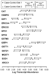
Figure 3. Two transcript expression cut-points best separate cancer from non-cancer. (one column width)
Using the modified Youden Index (J) method, for each of the 14 antioxidant, DNA repair and transcription factor genes, two cut-points were identified that best separated cancer from non-cancer (Supplementary Table 4). Cut-point levels are displayed in units of Log10 transformed target gene transcript abundance molecules per 106 ACTB transcript molecules. Arrows stemming from the points indicate the range of values with higher likelihood of cancer diagnosis compared to the ranges between the two cut-points, which are indicative of the range of values associated with lower likelihood of cancer diagnosis. Genes are listed in HUGO gene nomenclature format.
Figure 4
Figure 4. ROC analysis of the 14-gene composite lung cancer marker. (two column width)
ROC analysis was used to assess the ability of RTV to correctly classify each subject into the cancer or non-cancer group in the first case-control set (panel A), second set (panel B) or combined sets (panel C). AUC = Area Under the receiver Curve.
Figure 5
Figure 5. Lung cancer discrimination using Risk Test Value and age. (two column width)
Plotted is the multigene Risk Test Value (RTV) as a function of age (years) for the combined set of 89 bronchial epithelial cell samples. RTV x age = 420 gave the best discrimination between lung cancer case samples and controls.

"VSports手机版" References

    1. Alberg AJ, Samet JM. Epidemiology of lung cancer. Chest. 2003;123:21S–49S. - PubMed (VSports)
    1. Gloeckler Ries LA, Reichman ME, Lewis DR, Hankey BF, Edwards BK. Cancer survival and incidence from the Surveillance, Epidemiology, and End Results (SEER) program. Oncologist. 2003;8:541–52. - PubMed
    1. Doll R, Hill AB. A study of the aetiology of carcinoma of the lung. Br Med J. 1952;2:1271–86. - PMC - PubMed
    1. Shields PG. Molecular epidemiology of lung cancer. Ann Oncol. 1999;10 (Suppl 5):S7–11. - PubMed (V体育ios版)
    1. Ganti AK, Mulshine JL. Lung cancer screening. Oncologist. 2006;11:481–7. - PubMed

Publication types

MeSH terms