Skip to main page content
U.S. flag

An official website of the United States government

Dot gov

The . gov means it’s official. Federal government websites often end in . gov or VSports app下载. mil. Before sharing sensitive information, make sure you’re on a federal government site. .

Https

The site is secure. The https:// ensures that you are connecting to the official website and that any information you provide is encrypted and transmitted securely V体育官网. .

. 2009 Oct 14;29(41):13079-89.
doi: 10.1523/JNEUROSCI.3610-09.2009.

VSports手机版 - Protein phosphatase 1 regulates the histone code for long-term memory

Affiliations

Protein phosphatase 1 regulates the histone code for long-term memory

Kyoko Koshibu et al. J Neurosci. .

Abstract (V体育2025版)

Chromatin remodeling through histone posttranslational modifications (PTMs) and DNA methylation has recently been implicated in cognitive functions, but the mechanisms involved in such epigenetic regulation remain poorly understood. Here, we show that protein phosphatase 1 (PP1) is a critical regulator of chromatin remodeling in the mammalian brain that controls histone PTMs and gene transcription associated with long-term memory. Our data show that PP1 is present at the chromatin in brain cells and interacts with enzymes of the epigenetic machinery including HDAC1 (histone deacetylase 1) and histone demethylase JMJD2A (jumonji domain-containing protein 2A). The selective inhibition of the nuclear pool of PP1 in forebrain neurons in transgenic mice is shown to induce several histone PTMs that include not only phosphorylation but also acetylation and methylation. These PTMs are residue-specific and occur at the promoter of genes important for memory formation like CREB (cAMP response element-binding protein) and NF-kappaB (nuclear factor-kappaB). These histone PTMs further co-occur with selective binding of RNA polymerase II and altered gene transcription, and are associated with improved long-term memory for objects and space. Together, these findings reveal a novel mechanism for the epigenetic control of gene transcription and long-term memory in the adult brain that depends on PP1. VSports手机版.


Figures

Figure 1.
Figure 1.
Inducible and neuron-specific inhibition of nuclear PP1 in the adult mouse forebrain. a, Schematic representation of the NIPP1* fragment containing the PP1 inhibitory domain and nuclear localization signal (amino acids 143–224; red box) expressed as a transgene. b, Schematic of the transgenes used to express NIPP1* inducibly in forebrain neurons. NIPP1*-EGFP expression is induced by doxycycline (Dox) (On). c, RT-PCR examining NIPP1* and EGFP expression in cortex (Ctx), hippocampal formation (Hip), amygdala (Amy), and hypothalamus (Hyp) in adult mutant mice treated with dox or not. d, Immunohistochemical staining showing NIPP1*-EGFP colocalization with the neuronal marker NeuN in nuclei (NIPP1*-EGFP, green/NeuN, red), and nuclear counterstaining with hematoxylin (NIPP1*-EGFP, brown/Hemat, blue). Scale bar, 20 μm. e, Decreased PP1 activity in nuclear (n = 7) but not cytoplasmic (n = 5) hippocampal extracts from NIPP1*-EGFP mice compared with control littermates (nuclear, n = 11; cytoplasmic, n = 7). Nuclear PP1 activity, F(1,15) = 11.24, **p < 0.01; cytoplasmic activity, F(1,12) = 1.14, p = 0.31. Error bars indicate SEM.
Figure 2.
Figure 2.
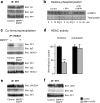
Nuclear PP1 interacts with the epigenetic machinery. a, Representative Western blot showing coimmunoprecipitation of H3 and PP1 using a PP1γ antibody in NIPP1*-EGFP mice and control littermates in the adult hippocampus. b, Direct dephosphorylation of histone H3 in vitro. Western blot showing S10 phosphorylation on H3 by protein kinase A (control) incubated with the catalytic subunit of PP1 (+ PP1), or with PP1 and NIPP1 (+ PP1, + NIPP1) for 0, 5, 10, or 30 min. Top panel, Anti-phospho H3S10; bottom panel, loading control (Coomassie blue gel staining after SDS-PAGE). c, Representative Western blot showing decreased PP1/HDAC1 interaction in HDAC1 immunoprecipitates in the presence of NIPP1* in vitro (top panel), and in the hippocampus of NIPP1*-EGFP mice (bottom panel). d, HDAC activity is decreased in nuclear (n = 7) but not cytoplasmic (n = 3) hippocampal extracts from NIPP1*-EGFP mice when compared with control littermates (nuclear, n = 7; cytoplasmic, n = 3); nuclear HDAC activity, F(1,12) = 32.02, ***p < 0.001; cytoplasmic, n.s., data are normalized to control littermates. Error bars indicate SEM. e, Representative Western blot showing decreased PP1/JMJD2A interaction in PP1 immunoprecipitates in the hippocampus of NIPP1*-EGFP mice. f, Representative Western blot showing no difference in PP1/LSD1 interaction in PP1 immunoprecipitates between control and mutant mice. Co-IP data are representative of at least three independent experiments.
Figure 3.
Figure 3.
Increase in specific histone PTMs by inhibition of nuclear PP1 and reversal by PP1γ overexpression in hippocampus. a, b, Representative Western blots (a) and corresponding quantitative analyses (b) of phosphorylation of H3T3 (control, n = 10; NIPP1*-EGFP, n = 9), H3S10 (control, n = 11; NIPP1*-EGFP, n = 10; F(1,19) = 8.57, p < 0.01), H3T11 (control, n = 10; NIPP1*-EGFP, n = 9), H3S28 (control, n = 10; NIPP1*-EGFP, n = 9), acetylation of H3K9 (control, n = 8; NIPP1*-EGFP, n = 9), H3K14 (control, n = 9; NIPP1*-EGFP, n = 12; F(1,19) = 7.59, p < 0.05), H4K5 (control, n = 6; NIPP1*-EGFP, n = 5; F(1,9) = 5.62, p < 0.05), H2B (control, n = 8; NIPP1*-EGFP, n = 8; F(1,14) = 7.32, p < 0.05), dimethylation of H3K4 (control, n = 5–8; NIPP1*-EGFP, n = 5–9), and trimethylation of H3K36 (control, n = 8; NIPP1*-EGFP, n = 11; F(1,17) = 7.91, p < 0.05) in nuclear extracts from the hippocampus (including CA1, CA3, and dentate gyrus) in behaviorally naive mice. Data are normalized to levels of total H1.0. c, d, Representative Western blots (c) and quantitative analyses (d) of histone PTMs in hippocampus injected with aCSF in control slices (control, n = 2–3 slices from 4 to 5 control mice), in NIPP1*-EGFP slices expressing injected with aCSF (NIPP1*, n = 2–3 slices from 3 to 4 NIPP1*-EGFP mice), in control slices injected with PP1γ-EGFP (PP1γ, n = 2–3 slices from 4 to 5 control mice), and in NIPP1*-EGFP slices injected with PP1γ-EGFP (PP1γ + NIPP1*, n = 2–3 slices from 4 to 5 NIPP1*-EGFP mice). Phosphorylation of H3S10, F(3,12) = 8.18, p < 0.01; LSD post hoc, control versus NIPP1*, p < 0.01; control versus PP1γ, p < 0.01; control versus PP1γ or PP1γ + NIPP1*, n.s; acetylation of H3K14, F(3,12) = 8.11, p < 0.01; LSD post hoc, control versus NIPP1*, p < 0.05; control versus PP1γ, p < 0.01; control versus PP1γ or PP1γ + NIPP1*, n.s; acetylation of H4K5, F(3,11) = 4.23, p < 0.01; LSD post hoc, control versus NIPP1*, p < 0.05; control versus PP1γ, p < 0.01; control versus PP1γ or PP1γ + NIPP1*, n.s; trimethylation of H3K36, F(3,12) = 13.40, p < 0.01; LSD post hoc, control versus NIPP1*, p < 0.05; control PP1γ, p < 0.01; control versus PP1γ or PP1γ + NIPP1*, n.s.; acetylation of H3K9 and dimethylation of H3K4, n.s. Data were first normalized to levels of total H1.0., and then to control slices injected with aCSF. Error bars indicate SEM. *p < 0.05, **p < 0.01.
Figure 4.
Figure 4.
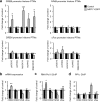
Promoter-specific histone PTMs and gene expression are altered by inhibition of nuclear PP1. a, ChIP assays showing histone PTMs in the promoter region of CREB, NF-κB, CREM, and c-Fos in the hippocampus (control, n = 3–6; NIPP1*-EGFP, n = 3–5): pH3S10 (CREB, t(5) = 2.78, p < 0.05; NF-κB, t(5) = 3.12, p < 0.05; CREM, n.s.; c-Fos, n.s.); AcH3K9 (CREB, n.s.; NF-κB, n.s.; CREM, t(5) = 5.38, p < 0.01; c-Fos, n.s.); AcH3K14 (CREB, t(7) = 6.58, p < 0.001; NF-κB, t(7) = 9.97, p < 0.001; CREM, n.s.; c-Fos, n.s.); AcH4K5 (CREB, t(5) = 3.04, p < 0.05; NF-κB, t(7) = 3.04, p < 0.05; CREM, n.s.; c-Fos, n.s.); 3MeH3K36 (CREB, t(9) = 2.42, p < 0.05; NF-κB, t(7) = 7.27, p < 0.001; CREM, n.s.; c-Fos, n.s.). b, Quantitative RT-PCR showing mRNA expression in control (n = 7–9) and NIPP1*-EGFP (n = 5–9) mice for CREB (F(1,16) = 9.84, p < 0.01), CREM (n.s.), NF-κB (F(1,10) = 10.97, p < 0.01), and c-Fos (n.s.). c, ChIP assays showing RNA Pol II occupancy in the promoter region of CREB, CREM, NF-κB, and c-Fos (control, n = 3–6; NIPP1*-EGFP, n = 3–4; CREB t(6) = 3.07, p < 0.05; CREM, n.s.; NF-κB, t(6) = 3.82, p < 0.05; c-Fos, n.s.). d, ChIP assays showing PP1γ binding to the promoter region of CREB, CREM, NF-κB, and c-Fos (control, n = 3–4; NIPP1*-EGFP, n = 3–4; CREB, t(6) = 6.26, p < 0.01; CREM, n.s.; NF-κB, t(6) = 3.44, p < 0.05; c-Fos, n.s.). Data are normalized to control littermates. Error bars indicate SEM. *p < 0.05, **p < 0.01, ***p < 0.001.
Figure 5.
Figure 5.
Object recognition and spatial memory are improved by inhibition of nuclear PP1. a, Object discrimination ratio in NIPP1*-EGFP (n = 8) and control littermates (n = 10) treated with dox (F(1,16) = 5.82, p < 0.05) (left panel) and in NIPP1*-EGFP (n = 7) and control littermates (n = 6) on/off dox (n.s.) 1 d after training (right panel). b, Object discrimination ratio in NIPP1*-EGFP (n = 8) and control littermates (n = 7) treated with dox (n.s.) (left panel) and in NIPP1*-EGFP (n = 6) and control littermates (n = 6) on/off dox (n.s.) 10 min after training (right panel). c, Left panels, Spatial memory on the water maze evaluated by the distance traveled in the target quadrant in NIPP1*-EGFP (n = 7) and control littermates (n = 8) treated with dox (F(1,13) = 5.39; p < 0.05) (left) and in NIPP1*-EGFP (n = 3) and control littermates (n = 6) on/off dox (n.s.) (right) 1 d after the end of training. Middle panels, Spatial memory measured by the number of platform crossings in NIPP1*-EGFP (n = 7) and control littermates treated with dox (n = 8) (F(1,13) = 12.05; p < 0.01) (left) and in NIPP1*-EGFP (n = 3) and control littermates (n = 6) on/off dox (n.s.) (right) 1 d after the end of training. Right panels, Spatial memory measured by time spent in target quadrant in NIPP1*-EGFP (n = 7) and control littermates treated with dox (n = 8) (F(1,13) = 5.93; p < 0.05) (left) and in NIPP1*-EGFP (n = 3) and control littermates (n = 6) on/off dox (right) 1 d after the end of training; behavioral data are representative of a minimum of two experiments on each task using independent groups of mice. Error bars indicate SEM. *p < 0.05, **p < 0.01.
Figure 6.
Figure 6.
Changes in histone PTMs after object recognition. a, ChIP analyses of H3S10 phosphorylation, H3K9, H3K14, and H4K5 acetylation, and H3K36 trimethylation at the CREB promoter in the hippocampus after novel object recognition (control naive and trained, n = 3–4; NIPP1*-EGFP naive and trained, n = 3–4). pH3S10, naive, control versus mutant, t(5) = 7.4, p < 0.01; trained, control versus mutant, t(5) = 4.3, p < 0.05; control, trained versus naive, t(5) = 5.87, p < 0.01; mutant, trained versus naive, t(5) = 4.44, p < 0.05; difference between slopes, p < 0.05. AcH3K9, naive, control versus mutant, n.s.; trained, control versus mutant, n.s.; control, trained versus naive, n.s; mutant, trained versus naive, t(5) = 4.91, p < 0.01; difference between slopes, p < 0.05. AcH3K14, naive, control versus mutant, t(4) = 8.58, p < 0.01; trained, control versus mutant, t(5) = 12.01, p < 0.001; control, trained versus naive, t(5) = 7.07, p < 0.01; mutant, trained versus naive, t(5) = 5.42, p < 0.05; difference between slopes, p < 0.05. AcH4K5, naive, control versus mutant, t(5) = 6.29, p < 0.01; trained, control versus mutant, t(5) = 3.42, p < 0.05; control, trained versus naive, n.s.; mutant, trained versus naive, t(5) = 5.22, p < 0.01; difference between slopes, p < 0.05. 3meH3K36, naive, control versus mutant, t(5) = 4.85, p < 0.01; trained, control versus mutant, t(4) = 10.06, p < 0.01; control, trained versus naive, n.s.; mutant, trained versus naive, t(5) = 5.45, p < 0.01; difference between slopes, p < 0.01. Data are normalized to control naive littermates. b, Quantitative RT-PCR showing CREB mRNA expression over time (control naive and trained, n = 5–8; NIPP1*-EGFP naive and trained, n = 5–8; naive, control vs mutant, F(1,8) = 20.76, p < 0.01; trained, control vs mutant, F(1,8) = 5.7, p < 0.05; control, trained vs naive, F(1,8) = 18.13, p < 0.01; mutant, trained vs naive, F(1,8) = 5.15, p < 0.05). Data are normalized to control naive littermates. c, Quantitative analysis (below) of the ratio between phosphorylated CREB and total CREB in the hippocampus in mutant and control mice; representative Western blots are shown above bar charts. β-Actin was used as an internal control and data were normalized to control littermates. Naive, control versus mutant, n.s.; trained, control versus mutant, F(1,6) = 6.54, p < 0.05; control, trained versus naive, n.s.; mutant, trained versus naive, F(1,8) = 8.64, p < 0.05. The asterisks indicate a significant difference between mutant and control mice; the hash signs a significant difference within the same treatment (mutant or control) group. Error bars indicate SEM. *p < 0.05, **p < 0.01, ***p < 0.001.
Figure 7.
Figure 7.
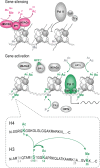
Model for a PP1-dependent histone code for the control of gene transcription. For gene silencing, PP1 binds to the chromatin where it dephosphorylates histones and negatively controls histone acetylation and methylation through association with HDACs and JMJD2A. These changes enhance chromatin condensation and prevent RNA polymerase II (Pol II), TATA box binding protein (TBP), and other transcription factors (TFs) from binding to the chromatin. For gene activation, PP1 is inhibited by endogenous inhibitors such as NIPP1* or targeting partners. This favors histone phosphorylation (specifically on H3S10), which is mediated by protein kinases (PKs) such as MSK1, ERK1, or PKA (Canettieri et al., 2003; Chwang et al., 2006, 2007). PP1 inhibition further leads to the dissociation of PP1 from HDACs, which reduces HDAC activity and thus increases histone acetylation (specifically on H3K14 and H4K5), most likely with the aid of HATs such as CBP (Alarcón et al., 2004; Korzus et al., 2004) and p300 (Oliveira et al., 2007). These changes are accompanied by an increase in methylation of H3K36, which is likely to result from a reduced interaction between PP1 and JMJD2A and a subsequent reduction in the activity of JMJD2A. Other members of the epigenetic machinery such as histone methyltransferases (HMTs) are presumably also involved. Together, these changes trigger chromatin decondensation and increase its accessibility to the transcriptional machinery. Bottom part, in gray, histone residues assessed for PTMs but not found to be differentially regulated; in green, histone PTMs, which depend on nuclear PP1. The thick arrow represents a well established cross talk between H3S10 phosphorylation and H3K14 acetylation in the context of memory formation (Chwang et al., 2006, 2007). The thin arrows illustrate potential cross talks suggested by the present data. For clarity, the cross talk between these residues and the acetylated H2B is omitted. Note that some of these cross talks have been reported in other model organisms (Kouzarides, 2007; Latham and Dent, 2007), but not in the adult mammalian brain.

VSports注册入口 - References

    1. Abel T, Zukin RS. Epigenetic targets of HDAC inhibition in neurodegenerative and psychiatric disorders. Curr Opin Pharmacol. 2008;8:57–64. - PMC - PubMed
    1. Alarcón JM, Malleret G, Touzani K, Vronskaya S, Ishii S, Kandel ER, Barco A. Chromatin acetylation, memory, and LTP are impaired in CBP+/− mice: a model for the cognitive deficit in Rubinstein-Taybi syndrome and its amelioration. Neuron. 2004;42:947–959. - PubMed
    1. Arthur JS. MSK activation and physiological roles. Front Biosci. 2008;13:5866–5879. - PubMed
    1. Bennett D. Transcriptional control by chromosome-associated protein phosphatase-1. Biochem Soc Trans. 2005;33:1444–1446. - PubMed
    1. Beullens M, Van Eynde A, Stalmans W, Bollen M. The isolation of novel inhibitory polypeptides of protein phosphatase 1 from bovine thymus nuclei. J Biol Chem. 1992;267:16538–16544. - PubMed

VSports最新版本 - Publication types

MeSH terms