V体育平台登录 - Skip to main page content
U.S. flag

An official website of the United States government

Dot gov

The . gov means it’s official. Federal government websites often end in . gov or . mil. Before sharing sensitive information, make sure you’re on a federal government site. VSports app下载.

Https

The site is secure. The https:// ensures that you are connecting to the official website and that any information you provide is encrypted and transmitted securely. V体育官网.

. 2010 Jan;159(1):1-10.
doi: 10.1111/j.1365-2249.2009.04031.x. Epub 2009 Oct 6.

"VSports" Laser microdissection-based analysis of cytokine balance in the kidneys of patients with lupus nephritis

Affiliations

"VSports在线直播" Laser microdissection-based analysis of cytokine balance in the kidneys of patients with lupus nephritis

Y Wang et al. Clin Exp Immunol. 2010 Jan.

"VSports手机版" Abstract

To determine the cytokine balance in patients with lupus nephritis (LN), we analysed kidney-infiltrating T cells. Renal biopsy samples from 15 systemic lupus erythematosus (SLE) patients were used. In accordance with the classification of International Society of Nephrology/Renal Pathology Society, they were categorized into Class III, Class III+V (Class III-predominant group, n = 4), Class IV, Class IV+V (Class IV-predominant group, n = 7) and Class V (n = 4) groups. The single-cell samples of both the glomelular and interstitial infiltrating cells were captured by laser-microdissection VSports手机版. The glomerular and interstitial infiltrating T cells produced interleukin (IL)-2, IL-4, IL-10, IL-13 and IL-17 cytokines in the Class III-predominant, Class IV-predominant and Class V groups. Interferon-gamma was detected only in the glomeruli of the Class III-predominant and Class V group samples. The expression level of IL-17 was correlated closely with clinical parameters such as haematuria, blood urea nitrogen level, SLE Disease Activity Index scores in both glomeruli and interstitium, urine protein level in glomeruli and serum creatinine and creatinine clearance levels in interstitium. This suggests that the glomerular infiltrating T cells might act as T helper type 1 (Th1), Th2 and Th17 cells while the interstitial infiltrating T cells, act as Th2 and Th17 cells in the Class III-predominant and Class V groups. In contrast, both the glomerular and interstitial infiltrating T cells might act as Th2 and Th17 cells in the Class IV-predominant group. The cytokine balances may be dependent upon the classification of renal pathology, and IL-17 might play a critical role in SLE development. .


Figures

Fig. 2
Fig. 2
(A) Targeted infiltrating cells selected and cut by laser microdissection (LMD). The glomeruli and interstitium areas of a single infiltrating cell (black arrows) were selected and dissected with a laser microbeam one by one. (B) Analysis of cytokine gene expression in lesions. (a) Detection of cytokines in the lesions of the renal biopsy specimens from the patients by nested polymerase chain reaction (PCR). Specific expression of interferon (IFN)-γ, interleukin (IL)-2, IL-4, IL-10, IL-13 and IL-17 were identified in the lesions of the glomeruli and interstitium areas from the renal biopsy specimens of the patients in the Class III-predominant groups (n = 4), Class IV-predominant groups (n = 7) and Class V groups (n = 4). M: molecular size marker; NC: negative control; PC: positive control cDNA clone. (b) Expression of IFN-γ, IL-2, IL-4, IL-10, IL-13 and IL-17 mRNAs in the glomeruli areas of the Class III-predominant (white bars), Class IV-predominant (hatched bars) and Class V (black bars) groups was analysed by nested reverse transcription–polymerase chain reaction (RT–PCR). (c) Expression of IFN-γ, IL-2, IL-4, IL-10, IL-13 and IL-17 mRNAs in the interstitium areas of the Class III-predominant (white bars), Class IV-predominant (hatched bars) and Class V (black bars) groups was analysed by nested RT–PCR (n.d. = not determined). The number of positive samples is shown as a percentage. Error bars represent ± standard error. P < 0·05, by Mann–Whitney U-test.
Fig. 1
Fig. 1
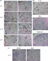
Detection of T cells in glomeruli and interstitium. (A) Stained interleukin (IL)-4, IL-10, IL-13 and IL-17 were observed in glomeruli and interstitium areas of the Class IV-predominant groups. Many IL-4 cells are observed prominently, mainly in the cells infiltrating into the glomeruli and interstitium areas, especially in intraglomerular infiltrating cells. There are only a few IL-4 cells in the tubular epithelial cells (TEC) (a, b). IL-10 and IL-13 cells are observed prominently in some of the cells infiltrating into the glomeruli and interstitium areas (c–f). Some stained IL-10 cells were observed in TEC (d). Many IL-17 cells are observed prominently, mainly in the cells infiltrating into the glomeruli, interstitium areas and TEC (g, h), especially in intraglomerular cells and TEC (g) (original magnification × 100 in a–h). (B) In the Class III-predominant group, some stained IL-17 cells were observed in TEC but almost no IL-17-positive cells were observed in glomeruli (a). Interferon (IFN)-γ cells were not observed in all the specimens (b). Normal rabbit immunoglobulin (Ig)G was stained as negative control (c) (original magnification × 100). (C) IL-17-positive cells were not observed in all the specimens of minor glomerular abnormalities (MGA) and minimal change nephrotic syndrome (MCNS) (a, b) (original magnification × 100).
Fig. 3
Fig. 3
Correlation between T helper type 1 (Th1), Th2 and Th17 cytokines and clinical and laboratory parameters in systemic lupus erythematosus (SLE). (A) Correlation between the levels of Th1 cytokine interleukin (IL)-2 and anti-double-strand (ds) DNA antibodies (a), 50% haemolytic unit of complement serum (CH50) (b), creatinine clearance (c) and chronicity index score (d) in glomeruli (black full line and points) and interstitium (black dashed line and grey points). (B) Correlation between the levels of Th2 cytokines–IL-4 and haematuria (a), blood urea nitrogen (b), serum creatinine (c) and chronicity index score (d) in glomeruli (black full line and points). Correlation between the levels of IL-10 and urine protein in interstitium (black dashed line and grey points). (C) Correlation between the levels of IL-13 and blood urea nitrogen (a), serum creatinine (b), 50% haemolytic unit of complement serum (CH50) (c), anti-ds DNA antibodies (d) and Activity Index scores (e) in interstitium. (D) Correlation between the levels of Th17 cytokine (IL-17), haematuria (a) and SLE Disease Activity Index (SLEDAI) scores (b) in glomeruli (black full line and points) and interstitium (black dashed line and grey points). Correlation between the level of IL-17 and SLEDAI scores (> 10) in glomeruli and interstitium (c). A simple linear regression analysis was used to evaluate the correlation between the two parameters, P < 0·05.

References

    1. Funauchi M, Ikoma S, Enomoto H, Horiuchi A. Decreased Th-1 like and increased Th-2 like cells in systemic lupus erythematosus. Scand J Rheumatol. 1998;27:219–24. - PubMed
    1. Viallard JF, Pellegrin JL, Ranchin V, et al. Th1 [IL-2, interferon-gammma (IFN-γ)] and Th2 (IL-10, IL-4) cytokine production by peripheral blood mononuclear cells (PBMC) from patients with systemic lupus erythematosus (SLE) Clin Exp Immunol. 1999;115:189–95. - PMC - PubMed
    1. Richaud-Patin Y, Alcocer-Verela J, Llorente L. High levels of TH2 cytokine gene expression in systemic lupus erythematosus. Rev Invest Clin. 1995;47:267–72. - PubMed
    1. Akahoshi M, Nakashima H, Tanaka Y, et al. Th1/Th2 balance of peripheral T helper cells in systemic lupus erytematosus. Arthritis Rheum. 1999;42:1644–8. - PubMed
    1. Masutani K, Akahoshi M, Tsuruya K, et al. Predominance of Th1 immune response in diffuse proliferative lupus nephritis. Arthritis Rheum. 2001;44:2097–106. - PubMed

V体育平台登录 - Publication types

MeSH terms