Skip to main page content
U.S. flag

An official website of the United States government

Dot gov

The VSports app下载. gov means it’s official. Federal government websites often end in . gov or . mil. Before sharing sensitive information, make sure you’re on a federal government site. .

Https

The site is secure. The https:// ensures that you are connecting to the official website and that any information you provide is encrypted and transmitted securely V体育官网. .

. 2009 Sep 22;106(38):16163-8.
doi: 10.1073/pnas.0903015106. Epub 2009 Sep 10.

Profiling protein thiol oxidation in tumor cells using sulfenic acid-specific antibodies

Affiliations

Profiling protein thiol oxidation in tumor cells using sulfenic acid-specific antibodies

Young Ho Seo et al. Proc Natl Acad Sci U S A. .

Abstract

Hydrogen peroxide (H2O2) functions as a second messenger that can activate cell proliferation through chemoselective oxidation of cysteine residues in signaling proteins. The connection between H2O2 signaling, thiol oxidation, and activation of growth pathways has emerged as fertile ground for the development of strategies for cancer treatment. Central to achieving this goal is the development of tools and assays that facilitate characterization of the molecular events associated with tumorigenesis and evaluation of patient response to therapy. Here we report on the development of an immunochemical method for detecting sulfenic acid, the initial oxidation product that results when a thiolate reacts with H2O2. For this approach, the sulfenic acid is derivatized with a chemical tag to generate a unique epitope for recognition. The elicited antibody is exquisitely specific, context-independent, and capable of visualizing sulfenic acid formation in cells. Applying this approach to several systems, including cancer cell lines, shows it can be used to monitor differences in thiol redox status and reveals a diverse pattern of sulfenic acid modifications across different subtypes of breast tumors VSports手机版. These studies demonstrate a general strategy for producing antibodies against a specific oxidation state of cysteine and show the utility of these reagents for profiling thiol oxidation associated with pathological conditions such as breast cancer. .


Conflict of interest statement (V体育官网)

Conflict of interest statement: The University of Michigan is presently negotiating with Assay Designs to commercialize this antibody along with the necessary reagents for labeling protein sulfenic acids.

Figures

Fig. 1.
Fig. 1.
Strategy for tagging and antibody-based detection of protein sulfenic acids. (A) Chemoselective alkylation of sulfenic acids by dimedone yields the synthetic epitope, which is recognized by the α-hapten antibody. (B) Structure of the hapten conjugate used to elicit sulfenic acid-specific antibodies. (C) GAPDH was incubated with combinations of hydrogen peroxide and dimedone as indicated; reaction products were analyzed by Western blot with the α-hapten antibody.
Fig. 2.
Fig. 2.
Immunofluorescence microscopy and Western blot showing sulfenic acid-modified proteins in HeLa cells. (A) HeLa cells were incubated in media containing 5 mM dimedone (top and middle) or DMSO (2.5% vol/vol) (bottom) for 1 h at 37 °C and analyzed for protein sulfenic acids (red). Nuclei were counterstained with DAPI (blue). (Scale bars, 60 μm.) (B) HeLa cells were untreated or treated with 1 mM H2O2 for 5, 15, or 30 min at 37 °C. At the end of each time point, cells were washed and incubated in media containing 5 mM dimedone for 15 min at 37 °C and then analyzed for protein sulfenic acids (red). Nuclei were counterstained with DAPI (blue). (C) Protein was isolated from cells incubated in media containing 2.5, 5, or 10 mM dimedone or DMSO (2.5% vol/vol) for 2 h at 37 °C. Sulfenic acid-modified proteins were analyzed by Western blot with the α-hapten antibody. Arrowhead, protein bands corresponding to sulfenic acid-modified actin, GAPDH and PrxI (see also SI Materials and Methods); asterisk, immunoreactive background band.
Fig. 3.
Fig. 3.
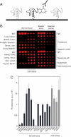
Detecting thiol oxidation using the protein microarray platform. (A) Strategy for detecting sulfenic acids on protein microarrays with the α-hapten antibody. (B) Profiling protein sulfenic acid levels in tissue lysates. The protein microarray was incubated in buffer containing 2 mM dimedone for 1 h and probed for sulfenic acids (red) with the α-hapten antibody. Negative (BSA and lysis buffer) and IgG (rabbit IgG spotted at 1.0 and 0.1 mg/mL) controls are also included on the array. (C) Analysis of sulfenic acid content in human tissues and mammalian cell lines. Chemifluorescence intensity of microarray spots is indicated with bars. Error bars, the standard deviation of three replicate spots.
Fig. 4.
Fig. 4.
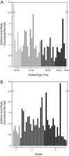
Comparison of sulfenic acid levels by protein microarrays from tumor and normal breast tissue lysates. The level of oxidation in each sample expressed as the ratio of sulfenic acid in breast tumor to the patient-matched normal and analyzed according to age (A) or histological grade (B). A ratio greater than one indicates that thiol oxidation is elevated in malignant tissue, relative to the paired sample. Chemifluorescence intensity of microarray spots is indicated with bars. The standard deviation for three replicate spots was less than ± 15%.
Fig. 5.
Fig. 5.
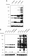
Profiling sulfenic acid modifications in breast cancer cells. (A) Comparative sulfenic acid analysis of matched breast cell lines HS578Bst (normal) and HS578T (carcinoma). HS578Bst and HS578T cells were cultured in media containing 5 mM dimedone or DMSO (2.5% vol/vol) for 2 h at 37 °C. After washing with PBS, cells were harvested in lysis buffer and sulfenic acid-modified proteins were analyzed by Western blot with the α-hapten antibody. Equal protein loading was verified by α-actin (bottom). Asterisks denote a background immunoreactive band. (B) Profiling sulfenic acid-modified proteins in breast cancer cell lines. BT20, BT474, MCF7, MDA-MB231, and MDA-MB468 cell lines were cultured in media containing 5 mM dimedone or DMSO (2.5% vol/vol) for 3 h at 37 °C. After washing with PBS, cells were harvested in lysis buffer and sulfenic acid-modified proteins were analyzed by Western blot using the α-hapten antibody. Equal protein loading was verified by α-actin (bottom). Asterisks denote a background immunoreactive band.

References

    1. Halliwell B, Gutteridge JMC. Free Radicals in Biology and Medicine. 4th Ed. New York: Oxford University Press; 2007. p. 851.
    1. Halliwell B. Oxidative stress and cancer: Have we moved forward? Biochem J. 2007;401:1–11. - PubMed
    1. Klaunig JE, Kamendulis LM. The role of oxidative stress in carcinogenesis. Annu Rev Pharmacol Toxicol. 2004;44:239–267. - PubMed
    1. Ragu S, et al. Oxygen metabolism and reactive oxygen species cause chromosomal rearrangements and cell death. Proc Natl Acad Sci USA. 2007;104:9747–9752. - PMC - PubMed
    1. Finkel T. Oxidant signals and oxidative stress. Curr Opin Cell Biol. 2003;15:247–254. - PubMed

Publication types

"VSports手机版" MeSH terms

LinkOut - more resources (V体育官网)