"VSports" Skip to main page content
U.S. flag

An official website of the United States government

Dot gov

The . gov means it’s official. Federal government websites often end in . gov or . mil VSports app下载. Before sharing sensitive information, make sure you’re on a federal government site. .

Https

The site is secure. The https:// ensures that you are connecting to the official website and that any information you provide is encrypted and transmitted securely V体育官网. .

. 2009 Nov 20;284(47):32522-32.
doi: 10.1074/jbc.M109.016139. Epub 2009 Sep 25.

"V体育ios版" Depletion of selenoprotein GPx4 in spermatocytes causes male infertility in mice

Affiliations

VSports - Depletion of selenoprotein GPx4 in spermatocytes causes male infertility in mice

Hirotaka Imai et al. J Biol Chem. .

"VSports手机版" Abstract

Phospholipid hydroperoxide glutathione peroxidase (GPx4) is an intracellular antioxidant enzyme that directly reduces peroxidized phospholipids. GPx4 is strongly expressed in the mitochondria of testis and spermatozoa. We previously found a significant decrease in the expression of GPx4 in spermatozoa from 30% of infertile human males diagnosed with oligoasthenozoospermia (Imai, H. , Suzuki, K. , Ishizaka, K VSports手机版. , Ichinose, S. , Oshima, H. , Okayasu, I. , Emoto, K. , Umeda, M. , and Nakagawa, Y. (2001) Biol. Reprod. 64, 674-683). To clarify whether defective GPx4 in spermatocytes causes male infertility, we established spermatocyte-specific GPx4 knock-out mice using a Cre-loxP system. All the spermatocyte-specific GPx4 knock-out male mice were found to be infertile despite normal plug formation after mating and displayed a significant decrease in the number of spermatozoa. Isolated epididymal GPx4-null spermatozoa could not fertilize oocytes in vitro. These spermatozoa showed significant reductions of forward motility and the mitochondrial membrane potential. These impairments were accompanied by the structural abnormality, such as a hairpin-like flagella bend at the midpiece and swelling of mitochondria in the spermatozoa. These results demonstrate that the depletion of GPx4 in spermatocytes causes severe abnormalities in spermatozoa. This may be one of the causes of male infertility in mice and humans. .


Figures (V体育平台登录)

FIGURE 1.
FIGURE 1.
Characterization of testes from 14-week-old spermatocyte-specific GPx4 knock-out mice (Tg(loxP):GPx4−/−). A and B, decreased testis size (A) and weight (B) in Tg(loxP):GPx4−/− mice. C, quantification of expression of GPx4 mRNA in several tissues of Tg(loxP-GPx4):GPx4−/− (white), Tg(loxP):GPx4−/− (black), and Tg(loxP):GPx4+/− (hatch) mice by quantitative real-time PCR. D, expression of GPx4, CuZn-SOD, Mn-SOD, and voltage-dependent anion channel protein in testes and epididymal spermatozoa of Tg(loxP-GPx4):GPx4−/− (1), Tg(loxP):GPx4−/− (2), and Tg(loxP):GPx4+/− (3) mice by immunoblot analysis with antibodies specific to each protein.
FIGURE 2.
FIGURE 2.
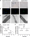
Morphological and functional abnormalities of cauda spermatozoa in spermatocyte-specific GPx4 knock-out mice (Tg(loxP):GPx4−/−). A–C, spermatozoa were flushed from the cauda epididymis of Tg(loxP-GPx4):GPx4−/− (left panel), Tg(loxP):GPx4−/− (center panel), and Tg(loxP):GPx4+/− mice (right panel) and analyzed by light and fluorescence microscopy. A, light microscopy showed that most epididymal spermatozoa of Tg(loxP):GPx4−/− mice exhibit a hairpin flagellar configuration (arrow). B, the mitochondrial membrane potential in epididymal spermatozoa was measured by incorporation of DiOC6. Fluorescence due to DiOC6 (green) and Hoechst 33258 (blue) was photographed under a fluorescence microscope. Scale bars, 50 μm. C, ultrastructure of mitochondria in the midpiece of spermatozoa by electron microscopy. Magnification, ×13,000. D and E, respiration capacity of cauda epididymal spermatozoa collected from Tg(loxP-GPx4):GPx4−/− (D) and Tg(loxP):GPx4−/− mice (E). Oxygen uptake by spermatozoa was measured polarographically in the presence of 10 mm malate and 10 mm pyruvate and 0.76 μm ADP. The rate of oxygen uptake in respiration states 4 (V4) and 3 (V3) is expressed as nanomoles of O2·ml/min/108 cells.
FIGURE 3.
FIGURE 3.
In vitro fertilization assay. The same number of spermatozoa collected from Tg(loxP-GPx4):GPx4−/−, Tg(loxP):GPx4−/−, and Tg(loxP):GPx4+/− mice (n = 3) were used in the assay. Phase-contrast microscopy of in vitro fertilization by spermatozoa from Tg(loxP-GPx4):GPx4−/− mice (left) and Tg(loxP):GPx4−/− mice (right) after 6- and 24-h incubation. Spermatozoa from Tg(loxP-GPx4):GPx4−/− and Tg(loxP):GPx4+/− mice were clearly able to inseminate eggs as shown by fertilized eggs at the two-cell stage (left bottom panel). Eggs incubated with spermatozoa from Tg(loxP):GPx4−/− mice showed no evidence of fertilization (bottom right panel). Scale bars, 50 μm. The development frequency of two-cell-stage embryos after 24 h was used as the measure of successful fertilization rate. No fertilization of spermatozoa from Tg(loxP):GPx4−/− mice was observed. All values are the means ± S.D.
FIGURE 4.
FIGURE 4.
The developmental progression of defects in the flagellar structure and dysfunction of the mitochondrial membrane potential in spermatozoa of spermatocyte-specific GPx4 knock-out mice (Tg(loxP):GPx4−/−). A–C, phase-contrast photomicrographs show the developmental progression of sperm defects in spermatocyte-specific GPx4 knock-out mice. Spermatozoa were flushed from seminiferous tubules (A), the caput epididymis (B), and cauda epididymis (C) from Tg(loxP):GPx4−/− mice observed by light microscopy. d–F, time course of the decrease of the mitochondrial membrane potential of GPx4-depleted cauda epididymal spermatozoa. Spermatozoa collected from cauda epididymis were cultivated for the indicated times, 0 h (D), 1 h (E), and 2 h (F), and stained with 10 μg/ml DiOC6 (green) and 1 μg/ml Hoechst 33258 (blue) for 20 min. Fluorescence from DiOC6 and Hoechst 33258 was photographed under a fluorescence microscope. Scale bars, 50 μm.
FIGURE 5.
FIGURE 5.
Histopathological observation of testes from spermatocyte-specific GPx4 knock-out mice (Tg(loxP):GPx4−/−). A and B, histological observation of testes. Testis sections from Tg(loxP-GPx4):GPx4−/−, Tg(loxP):GPx4−/−, and Tg(loxP):GPx4+/− mice were stained with hematoxylin and eosin. A, scale bars, 200 μm. B, scale bars, 50 μm. C, analysis of germ cell DNA content in testes from Tg(loxP-GPx4):GPx4−/−, Tg(loxP):GPx4−/−, and Tg(loxP):GPx4+/− mice by flow cytometry. 1N represents haploid cells, 2N represents diploid cells, and 4N represents tetraploid cells. S-phase (S-Ph) represents spermatogonial cells, and preleptotene spermatocytes synthesizing DNA. D, immunohistochemical analysis of the distribution of stage-specific germ cell. Testis sections from Tg(loxP-GPx4):GPx4−/− and Tg(loxP):GPx4−/− mice were stained with EE2 pAb, BC7 pAb, and anti-calmegin pAb to visualize spermatogonia (EE2), early spermatocytes (BC7), and late spermatocytes and spermatids (Calmegin). Arrows indicate the location of spermatogonia, early spermatocytes, and late spermatocytes in each panel. Scale bars, 50 μm.
FIGURE 6.
FIGURE 6.
Detection of hydroperoxide and caspase 3 activation in germ cells of spermatocyte-specific GPx4 knock-out mice (Tg(loxP):GPx4−/−). A, flow cytometric analysis of intracellular hydroperoxides in germ cells of Tg(loxP-GPx4):GPx4−/− (left panel), Tg(loxP):GPx4−/− (center panel), and Tg(loxP):GPx4+/− mice (right panel). To assess the levels of intracellular peroxides, flow cytometric analysis was performed with the fluorescent probe, dihydrorhodamine (DHR). The intensity of fluorescence from DHR of cells was quantified by flow cytometry and is plotted on a logarithmic scale, in arbitrary units, against the number of cells. B, detection of activated caspase 3, an apoptotic cell death regulator, in testis of Tg(loxP-GPx4):GPx4−/−, Tg(loxP):GPx4−/−, and Tg(loxP):GPx4+/− mice. Testis sections from each mouse were stained with Cy3-conjugated anti-activated caspase 3 pAb. Scale bars, 50 μm.

V体育官网 - References

    1. Sharma R. K., Agarwal A. (1996) Urology 48, 835–850 - PubMed
    1. Lipschultz L. I., Howards S. S. (1983) in Infertility in the Male (Lipschultz L. I., Howards S. S. eds) pp. 187– 206, Churchill Livingstone, New York
    1. Hull M. G., Glazener C. M., Kelly N. J., Conway D. J., Foster P. A., Hinton R. A., Coulson C., Lambert P. A., Watt E. M., Desai K. M. (1985) Br. Med. J. 291, 1693–1697 - PMC - PubMed
    1. Nishimune Y., Tanaka H. (2006) J. Androl. 27, 326–334 - "V体育安卓版" PubMed
    1. Imai H., Nakagawa Y. (2003) Free Radic. Biol. Med. 34, 145–169 - PubMed

Publication types

MeSH terms

Substances