Skip to main page content
U.S. flag

An official website of the United States government

Dot gov

The . gov means it’s official. Federal government websites often end in . gov or . mil. Before sharing sensitive information, make sure you’re on a federal government site. VSports app下载.

Https

The site is secure V体育官网. The https:// ensures that you are connecting to the official website and that any information you provide is encrypted and transmitted securely. .

. 2009 Oct;119(10):2925-41.
doi: 10.1172/JCI38857. Epub 2009 Sep 8.

"VSports最新版本" Calcium/calmodulin-dependent protein kinase II links ER stress with Fas and mitochondrial apoptosis pathways

Affiliations

Calcium/calmodulin-dependent protein kinase II links ER stress with Fas and mitochondrial apoptosis pathways

Jenelle M Timmins et al. J Clin Invest. 2009 Oct.

Abstract

ER stress-induced apoptosis is implicated in various pathological conditions, but the mechanisms linking ER stress-mediated signaling to downstream apoptotic pathways remain unclear. Using human and mouse cell culture and in vivo mouse models of ER stress-induced apoptosis, we have shown that cytosolic calcium resulting from ER stress induces expression of the Fas death receptor through a pathway involving calcium/calmodulin-dependent protein kinase IIgamma (CaMKIIgamma) and JNK. Remarkably, CaMKIIgamma was also responsible for processes involved in mitochondrial-dependent apoptosis, including release of mitochondrial cytochrome c and loss of mitochondrial membrane potential. CaMKII-dependent apoptosis was also observed in a number of cultured human and mouse cells relevant to ER stress-induced pathology, including cultured macrophages, endothelial cells, and neuronal cells subjected to proapoptotic ER stress. Moreover, WT mice subjected to systemic ER stress showed evidence of macrophage mitochondrial dysfunction and apoptosis, renal epithelial cell apoptosis, and renal dysfunction, and these effects were markedly reduced in CaMKIIgamma-deficient mice. These data support an integrated model in which CaMKII serves as a unifying link between ER stress and the Fas and mitochondrial apoptotic pathways. Our study also revealed what we believe to be a novel proapoptotic function for CaMKII, namely, promotion of mitochondrial calcium uptake. These findings raise the possibility that CaMKII inhibitors could be useful in preventing apoptosis in pathological settings involving ER stress-induced apoptosis VSports手机版. .


"V体育官网入口" Figures
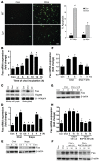
Figure 1
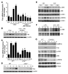
Figure 1. Proapoptotic Fas is induced by ER stress in a calcium-dependent manner.
(A) Peritoneal macrophages from WT or Lpr mice were incubated for 14 hours under control (Con) or cholesterol-loading (Chol) conditions. Mid- and late-stage apoptosis were assayed using Alexa Fluor 488–conjugated annexin V (green) and PI (orange-red), respectively. Scale bar: 20 μm. For each group, 3 fields were quantified and expressed as a percentage of annexin/PI-positive cells. Mϕ, macrophage. (B) Macrophages from WT mice were incubated under cholesterol-loading conditions for the indicated times. Fas mRNA, measured using quantitative PCR, is expressed relative to internal control mRNA. (C) Macrophages were incubated for 8 hours under control, cholesterol-loading, or cholesteryl ester–loading (CE) conditions and then assayed for total and cell surface Fas protein by immunoblot following biotinylation of cell surface proteins. Hsp90 was used as a cytosolic marker, and β1 integrin as a cell surface marker and loading control. (D and E) Macrophages were incubated under control or cholesterol-loading conditions or with 0.25 μM thapsigargin (Thaps) and then assayed for Fas mRNA and protein. (F and G) Macrophages were incubated under control or cholesterol-loading conditions in the absence or presence of the cholesterol trafficking inhibitor U18666A (U18) after 1 hour pretreatment with the compound, and then assayed for Fas mRNA and protein. (H and I) Macrophages were incubated under control or cholesterol-loading conditions with BAPTA-AM or equivalent volumes of vehicle (Veh) control after 1 hour pretreatment, and then assayed for Fas mRNA and protein. Differing symbols indicate P < 0.01; identical symbols indicate differences that are not significant.
Figure 2
Figure 2. Cholesterol loading leads to sustained activation of CaMKII.
(A) CaMKII activity was assayed in individual wells of macrophages incubated under cholesterol-loading conditions for the indicated times. The asterisk indicates CaMKII activity from cells loaded with cholesterol for 4 hours in the presence of 10 μM AIP-II (EGTA was included during the assay). (B) CaMKII activity was assayed in triplicate wells of macrophages incubated under control or cholesterol-loading conditions for 6 hours. Differing symbols indicate P = 0.001. (C) Top: Macrophages were incubated under control or cholesterol-loading conditions with BAPTA-AM or vehicle after 1 hour pretreatment, and lysates were then immunoblotted for phospho-CaMKII, total CaMKII, and β-actin. Bottom: As above, except ER stress was induced with 3 μg/ml tunicamycin (Tun). (D) Macrophages were incubated under control or cholesterol-loading conditions with or without 10 μM AIP-II after 1 hour pretreatment, and lysates were then immunoblotted for phospho-CaMKII, total CaMKII, and β-actin. (E and F) As in D, except that (E) ER stress was induced with 0.25 μM thapsigargin, or (F) 10 μM KN93 was used as the CaMKII inhibitor, with 10 μM KN92 as the inactive analog. (G) In 2 independent experiments, macrophages were incubated under control or cholesterol-loading conditions. CaMKII was then immunoprecipitated from cell lysates using anti-CaMKII or control IgG (bottom). The immunoprecipitates were then blotted for oxidized CaMKII using anti–Ox-CaMKII antiserum and total CaMKII.
Figure 3
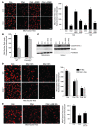
Figure 3. Induction of Fas and apoptosis by ER stress involves CaMKII.
(A) Macrophages were incubated for 8 hours under cholesterol-loading conditions with or without KN93 or KN92 after 1 hour pretreatment, and then assayed for Fas mRNA. (B) RNA from peritoneal macrophages from 4 separate mice and from mouse brain, along with water control, were probed for the indicated CaMKII isoform mRNAs by RT-PCR. (C) Macrophages were transfected with 3 different CaMKIIγ siRNA constructs. After 72 hours, the cells were incubated for 8 hours under cholesterol-loading conditions and then assayed for CaMKIIγ mRNA and total Fas protein. (D) Macrophages were transfected with the 3 siRNA constructs in C, incubated for 30 hours with 0.25 μM thapsigargin and 25 μg/ml of the SRA ligand fucoidan, and then assayed for apoptosis. (E) Macrophages from WT or Camk2g–/– mice were incubated under control or cholesterol-loading conditions for 12 hours and then assayed for apoptosis. Scale bar: 20 μm. (F) Human aortic endothelial cells were incubated for 24 hours with thapsigargin (1 μM) with or without KN93 or KN92 after 1 hour pretreatment, and then assayed for apoptosis. (G) PC12 cells were incubated for 24 hours with 100 μM 6-OHDA with or without 10 μM AIP-II after 1 hour pretreatment, and then assayed for cell viability (percentage of viable cells compared with those in cultures not treated with 6-OHDA). Differing symbols indicate P < 0.01; identical symbols indicate differences that are not significant.
Figure 4
Figure 4. JNK mediates the proapoptotic role of CaMKII in ER-stressed macrophages.
(A and B) Macrophages were incubated under control or cholesterol-loading conditions with or without 10 μM of the JNK inhibitor SP600125 after 1 hour pretreatment, and then assayed for Fas mRNA (A) and protein (B). (C and D) Macrophages from WT or Jnk2–/– mice were incubated under control or cholesterol-loading conditions and then assayed for Fas mRNA (C) and protein (D). (E) Macrophages were incubated under control or cholesterol-loading conditions for 4 hours with or without 10 μM AIP-II after 1 hour pretreatment. One aliquot of the cell extracts was immunoblotted for phospho-JNK, total JNK, and β-actin, and another was immunoblotted for phospho-MKK4 and β-actin. (F) Macrophages were incubated with 100 ng/ml LPS for the indicated times with or without 10 μM AIP-II after 1 hour pretreatment. Phospho-JNK and total JNK were then assayed by immunoblot. (G) Macrophages from WT or Camk2g–/– mice were incubated under control or cholesterol-loading conditions and then immunoblotted for CaMKII, Fas, phospho-JNK, JNK, phospho-MKK4, MKK4, and β-actin. Differing symbols indicate P < 0.01; identical symbols indicate differences that are not significant.
Figure 5
Figure 5. Release of mitochondrial cytochrome c and Δψm in cholesterol-loaded macrophages is dependent on CaMKII.
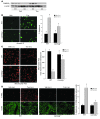
(A) Macrophages were incubated under control or cholesterol-loading conditions for 12 or 16 hours in the absence or presence of 10 μM KN93 or KN92 after 1 hour pretreatment. Cells were then stained with MitoTracker Red and examined by confocal fluorescence microscopy. Mean fluorescence intensity per cell is also shown (n = 100 cells). (B) Macrophages from WT or Camk2g–/– mice were incubated under control or cholesterol-loading conditions for 12 hours and then assayed for MitoTracker Red staining. (C) Macrophages were treated for 8 hours as in A. Mitochondrial and cytosolic fractions were assayed for cytochrome c, tubulin (cytosolic marker), and prohibitin (mitochondrial marker). (D) Macrophages from WT or Stat1–/– mice were incubated under control or cholesterol-loading conditions for 12 or 16 hours and then stained with MitoTracker Red. (E) Macrophages were incubated under control or cholesterol-loading conditions for 16 hours in the absence or presence of 10 μM of the JNK inhibitor SP600125 after 1 hour pretreatment, and then stained with MitoTracker Red. Scale bars: 20 μm (A); 10 μm (D and E). Differing symbols indicate P < 0.01; identical symbols indicate differences that are not significant.
Figure 6
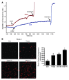
Figure 6. ER stress in macrophages leads to the accumulation of calcium in the mitochondria.
(A) Macrophages were plated on 25-mm coverslips and grown to confluency. The cells were incubated under control or cholesterol-loading conditions for 1 hour and stained with 5 μM Fura-2 for 30 minutes. The cells were then rinsed in medium and examined by fluorescence microscopy, where calcium fluorescence was measured every second. FCCP (1 μM), a mitochondrial uncoupler, and ionomycin (10 μM) were added at the indicated times to release mitochondrial and overall intracellular calcium stores, respectively. (B) Macrophages were treated with medium alone as control, with 0.25 μM thapsigargin for 1 hour, or under cholesterol-loading conditions for 2 or 4 hours. At the end of each incubation, 10 μM Rhod-2, a mitochondrial-specific fluorescent calcium dye, was added to the media, and the cells were incubated on ice for 1 hour at 4°C. The cells were then washed and incubated for an additional 5 hours with thapsigargin or under cholesterol-loading conditions, such that total incubation time for thapsigargin was 5 hours and for cholesterol 5 or 7 hours. At the end of the incubation period, the cells were visualized using confocal microscopy and imaged as described in Methods. Scale bar: 10 μm. Fluorescence intensity for approximately 100 cells was measured for each treatment group. Differing symbols indicate P < 0.01; identical symbols indicate differences that are not significant.
Figure 7
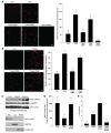
Figure 7. ER stress–induced mitochondrial calcium uptake is dependent on CaMKII.
(A and B) Macrophages were incubated under control or cholesterol-loading conditions (A) or with 0.25 mM thapsigargin as the ER stressor (B) for 7 hours, in the absence or presence of 10 μM KN93, KN92, or RU360 after 1 hour pretreatment. Rhod-2 labeling and quantification were then carried out as described in Figure 5. Scale bars: 10 μm. (C) Macrophages were incubated under control or cholesterol-loading conditions for the indicated times. Cytosol-free mitochondria were isolated and immunoblotted for total CaMKII, phospho-CaMKII, and cytochrome c oxidase (Cyto ox’ase). Mitochondria (Mit), cytosol (Cyt), and ER-plasma membrane (ER PM) fractions — immunoblotted for the mitochondrial marker cytochrome c oxidase, the cytosol marker GAPDH, and the ER marker calreticulin — are also shown. (D) Macrophages were incubated under control or cholesterol-loading conditions for 12 hours with or without 10 μM RU360 after 1 hour pretreatment, and then stained with MitoTracker Red. (E) Macrophages were incubated for 20 hours with medium alone as control or with medium containing 0.25 μM thapsigargin; thapsigargin and 50 μg/ml acetyl-LDL (AcLDL); or thapsigargin, acetyl-LDL, and RU360. Apoptosis was then assayed quantified. Differing symbols indicate P < 0.01; identical symbols indicate differences that are not significant.
Figure 8
Figure 8. Importance of CaMKIIγ in macrophage apoptosis and Δψm in ER-stressed mice.
(AC) WT and Camk2g–/– mice were injected i.v. with tunicamycin or vehicle control. Peritoneal macrophages were harvested 24 hours later and then assayed for CaMKIIγ protein by immunoblot analysis (A), for apoptosis by annexin V staining (B), or for mitochondrial membrane potential by MitoTracker Red staining (C). Scale bars: 10 μm. (D) WT and Camk2g–/– mice were injected i.p. with 1 mg/kg tunicamycin or vehicle control. The spleens were harvested 48 hours later and stained using TUNEL (apoptosis) or anti-CD68 (macrophages). Scale bar: 20 μm. Quantification of TUNEL-positive cells among CD68-positive cells from 3 mice per group is shown. Differing symbols indicate P < 0.01; identical symbols indicate differences that are not significant.
Figure 9
Figure 9. Importance of CaMKIIγ in renal tubular epithelial cell apoptosis and renal function in ER-stressed mice.
WT and Camk2g–/– mice were injected i.p. with 1 mg/kg tunicamycin or vehicle control. (A) The kidneys were harvested 48 hours later and stained using TUNEL (apoptosis) or DAPI (nuclei). Scale bar: 20 μm. Quantification of TUNEL-positive cells from 3 mice per group is shown. (B) Serum creatinine levels and urine albumin levels (normalized to urine creatinine) were determined for all groups of mice. Differing symbols indicate P < 0.01 (A) or P < 0.05 (B); identical symbols indicate differences that are not significant.
Figure 10
Figure 10. Schematic of calcium-CaMKII–mediated events leading to ER stress–induced macrophage apoptosis.
ER stress depletes the calcium stores within the ER lumen. Calcium subsequently accumulates in the cytoplasm and activates CaMKII. CaMKII activation, which is sustained through autophosphorylation and possibly ROS-mediated oxidation, enables apoptosis through at least 3 pathways: JNK-mediated Fas induction; promotion of mitochondrial calcium uptake, followed by mitochondrial membrane permeabilization (MMP), release of apoptogens, and Δψm; and activation of STAT1, a proapoptotic signal transducer. See text for details.

References

    1. Welihinda A.A., Tirasophon W., Kaufman R.J. The cellular response to protein misfolding in the endoplasmic reticulum. Gene Expr. 1999;7:293–300. - PMC - PubMed
    1. Ron D. Translational control in the endoplasmic reticulum stress response. J. Clin. Invest. 2002;110:1383–1388. - PMC - PubMed
    1. Feng B., et al. The endoplasmic reticulum is the site of cholesterol-induced cytotoxicity in macrophages. Nat. Cell Biol. 2003;5:781–792. doi: 10.1038/ncb1035. - "VSports" DOI - PubMed
    1. Marciniak S.J., et al. CHOP induces death by promoting protein synthesis and oxidation in the stressed endoplasmic reticulum. Genes Dev. 2004;18:3066–3077. doi: 10.1101/gad.1250704. - DOI - PMC - PubMed
    1. Szegezdi E., Logue S.E., Gorman A.M., Samali A. Mediators of endoplasmic reticulum stress-induced apoptosis. EMBO Rep. 2006;7:880–885. doi: 10.1038/sj.embor.7400779. - DOI - PMC - PubMed

Publication types

MeSH terms

"V体育官网入口" LinkOut - more resources