Skip to main page content
U.S. flag

An official website of the United States government

Dot gov

The . gov means it’s official. Federal government websites often end in . gov or VSports app下载. mil. Before sharing sensitive information, make sure you’re on a federal government site. .

Https

The site is secure. The https:// ensures that you are connecting to the official website and that any information you provide is encrypted and transmitted securely V体育官网. .

. 2009 Sep 15;183(6):3848-57.
doi: 10.4049/jimmunol.0901085. Epub 2009 Aug 26.

"VSports" Specific requirements for Vgamma9Vdelta2 T cell stimulation by a natural adenylated phosphoantigen

Affiliations

Specific requirements for Vgamma9Vdelta2 T cell stimulation by a natural adenylated phosphoantigen

VSports最新版本 - Pierre Vantourout et al. J Immunol. .

Abstract

Human Vgamma9Vdelta2 T lymphocytes recognize phosphorylated alkyl Ags. Isopentenyl pyrophosphate (IPP) was previously proposed as the main Ag responsible for Vgamma9Vdelta2 T cell activation by cancer cells. However, triphosphoric acid 1-adenosin-5'-yl ester 3-(3-methylbut-3-enyl) ester (ApppI), a metabolite in which the isopentenyl moiety is linked to ATP, was reported in cells activated with aminobisphosphonates. The contribution of this compound to tumor-stimulatory activity was thus examined. ApppI induces selective expansion of Vgamma9Vdelta2 T cells from PBMCs. In the absence of APCs, however, ApppI has little stimulatory activity on Vgamma9Vdelta2 T cells, and optimal activation with ApppI requires addition of a nucleotide pyrophosphatase releasing IPP plus AMP. Thus, ApppI has no intrinsic stimulatory activity VSports手机版. Nevertheless, stimulation by ApppI is strengthened by the presence of APCs. Moreover, in contrast to IPP, ApppI can be efficiently pulsed on dendritic cells as well as on nonprofessional APCs. Pulsed APCs display stable and phosphatase-resistant stimulatory activity, indicative of Ag modification. HPLC analysis of tumor cell extracts indicates that latent phosphoantigenic activity is stored intracellularly in the Vgamma9Vdelta2 cell-sensitive tumor Daudi and can be activated by a nucleotide pyrophosphatase activity. The presence of ApppI in Daudi cell extracts was demonstrated by mass spectrometry. Nucleotidic Ags such as ApppI are thus a storage form of phosphoantigen which may represent a major source of phosphoantigenic activity in tumor cells. The unique properties of ApppI may be important for the design of Ags used in anticancer immunotherapeutic protocols using Vgamma9Vdelta2 cells. .


Figures

Figure 1
Figure 1. Structure of ApppI and IPP
NPP and Apyr indicate the cleavage sites by nucleotide pyrophosphatase and apyrase.
Figure 2
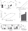
Figure 2. Activation of Vγ9Vδ2 T cells by ApppI
(A,B) PBMCs were stimulated with phosphoantigens and IL-2 before staining for CD3 and TCR Vδ2 expression. (A) PBMC staining before and after in vitro culture (19 days) with ApppI (5μM). Numbers indicate the percentage of cells in each quadrant. (B) Comparative outgrowth of Vδ2 T cell 1 week after PBMC stimulation with IPP or ApppI plus IL-2. (C) Intracellular IFN-γ staining of Vδ2+ cells following PBMC stimulation with IPP and ApppI. (D) A Vγ9Vδ2 T cell line was stimulated with the indicated antigens in the presence of anti-CD107a-PE antibody to measure the activation of the cytolysis machinery. Cells were then washed and stained for TCRVδ2. In dot plots, numbers indicate the percentage of CD107a+ cells among Vδ2+ cells after stimulation with antigens at 100μM. The dose-response curve is shown on the right. Data are representative ≥10 experiments. (E) PBMCs were stimulated in the presence of IPP (10μM) plus ApppI and stained as in C. (C,E) Results are means of triplicate cultures +/− SD.
Figure 3
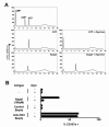
Figure 3. Effect of apyrase on ApppI and ApppI-induced lymphocyte response
(A) Individual nucleotides or nucleotide mixtures were either untreated or treated with apyrase before analysis by anion exchange chromatography to assess their sensitivity to the enzyme. (B) Vγ9Vδ2 T cells were stimulated with ApppI, anti-CD3 antibody or control IgG1-coated beads, or control medium in the presence or absence of apyrase, and the expression of CD107a was monitored as in Fig. 2D. Data are means of triplicate stimulation experiments +/− SD.
Figure 4
Figure 4. Effect of nucleotide pyrophosphatase on ApppI and ApppI-induced lymphocyte response
(A) Individual nucleotides or nucleotide mixtures were either untreated or treated with NPP before HPLC analysis as in Fig. 3A. (B) Vγ9Vδ2 T cells were stimulated with ApppI in the presence or absence of NPP and the expression of CD107a was monitored as in Fig. 2D. (C) The effect of NPP and its inhibitor PPADS was tested on lymphocyte stimulations by ApppI or anti-CD3 and monitored as in B. Data (B,C) are means of triplicate stimulation experiments +/− SD. *: p≤0.001; NS: non-significant (2-tailed Student test).
Figure 5
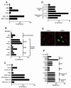
Figure 5. Effect of antigen presentation on ApppI response
Vγ9Vδ2 T cells were stimulated as indicated and CD107a expression was monitored as in Fig. 2D. Data are means of triplicate stimulation experiments +/− SD. (A) Stimulation with IPP, ApppI, RPMI-8226 cells (1:1 ratio) or RPMI-8226 plus phosphoantigens. (B) Indicated tumor cell lines were pulsed overnight with the indicated antigens before co-culture with Vγ9Vδ2 cells. (C) Daudi and K562 cells were treated overnight with the indicated compounds, washed and used as stimulators for Vγ9Vδ2 T cells. Lova: lovastatin (20μM); ApppI: 20μM; Pam: pamidronate (50μM). (D) RPMI-8226 cells were pulsed with ApppI (2h, 10μM) either at 37°C (Control), 4°C, or at 37°C after a 2h-pre-treatment with cytochalasin D (10μM), and washed extensively before addition of Vγ9Vδ2 T cells. (E) Vγ9Vδ2 T cells (surface-stained with anti-d2-FITC, green) were kept in contact for 20min at 37°C with K562 cells loaded with hexidium iodide (red) and loaded or not with ApppI. TCR aggregation was analyzed by confocal microscopy. (F) Tumor cells were pulsed overnight with pamidronate (50μM) or ApppI (20μM) or left untreated, washed, and polyclonal Vγ9Vδ2 T cells were added along with the indicated enzymes (Apyr: Apyrase, 0.2U/ml, NPP: Nucleotide Pyrophosphatase, 0.02U/ml).
Figure 6
Figure 6. Pulsing of phosphoantigens on dendritic cells
(A) DC were derived from CD14+ PBMCs (iDC) and maturation was induced by 48h LPS stimulation (mDC). They were subsequently pulsed with IPP or ApppI, extensively washed and co-cultured with Vγ9Vδ2 cells. T cell activation was monitored by CD107a expression. in Vδ2+ cells. (B) Representative pattern of CD83 and CD86 expression on DC populations before and after LPS treatment.
Figure 7
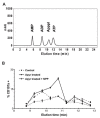
Figure 7. Daudi cells produce endogenous nucleotidic phosphoantigens
(A) Elution profile of AMP, ADP, ATP and ApppI. (B) Fractions eluted from Daudi cytosolic extracts were either untreated (control) or treated with apyrase. This was then denatured by heating and samples were assayed for Vγ9Vδ2 T cell stimulation with or without adding exogenous NPP. Data are means of triplicate stimulations +/− SD.
Figure 8
Figure 8. Detection of ApppI in Daudi cell extracts by mass spectrometry
(A) Nano ESI-ITMS analysis of synthetic ApppI. Top panel: full scan MS, showing the pseudo molecular ion signal of ApppI corresponding to ([M-H]). Middle panel: MS2 spectrum of m/z 574. Bottom panel: MS3 spectrum resulting from the fragmentation of the m/z 408 ion. (B) Nano ESI-ITMS analysis of biological extract corresponding to HPLC fraction eluting between 10.5 and 11 min (cf. Fig. 7A). Top Panel: MS2 spectrum of an m/z 574 ion from full scan, showing the characteristic m/z 408 ion. Bottom: spectrum resulting from the fragmentation of the m/z 408 ion showing a pattern similar to that of synthetic ApppI.

References

    1. Chen ZW, Letvin NL. Vgamma2Vdelta2+ T cells and anti-microbial immune responses. Microbes Infect. 2003;5:491–498. - PMC - PubMed
    1. Hayday AC. [gamma][delta] cells: a right time and a right place for a conserved third way of protection. Annu Rev Immunol. 2000;18:975–1026. - PubMed
    1. Kabelitz D, Wesch D, Pitters E, Zoller M. Potential of human gammadelta T lymphocytes for immunotherapy of cancer. Int J Cancer. 2004;112:727–732. - VSports - PubMed
    1. Dieli F, Vermijlen D, Fulfaro F, Caccamo N, Meraviglia S, Cicero G, Roberts A, Buccheri S, D’Asaro M, Gebbia N, Salerno A, Eberl M, Hayday AC. Targeting human {gamma}delta} T cells with zoledronate and interleukin-2 for immunotherapy of hormone-refractory prostate cancer. Cancer Res. 2007;67:7450–7457. - PMC - PubMed
    1. Constant P, Davodeau F, Peyrat MA, Poquet Y, Puzo G, Bonneville M, Fournie JJ. Stimulation of human gamma delta T cells by nonpeptidic mycobacterial ligands. Science. 1994;264:267–270. - VSports app下载 - PubMed

"VSports最新版本" Publication types

"VSports app下载" MeSH terms

Substances