<style draggable="OwG2kSGr"><noframes date-time="frry8GZ"><code dropzone="wgs8F"></code> Skip to main page content
U.S. flag

An official website of the United States government

Dot gov

The . gov means it’s official. Federal government websites often end in VSports app下载. gov or . mil. Before sharing sensitive information, make sure you’re on a federal government site. .

Https

The site is secure. The https:// ensures that you are connecting to the official website and that any information you provide is encrypted and transmitted securely V体育官网. .

. 2009 Aug 24;186(4):571-87.
doi: 10.1083/jcb.200812176.

Cortactin regulates cofilin and N-WASp activities to control the stages of invadopodium assembly and maturation

Affiliations

Cortactin regulates cofilin and N-WASp activities to control the stages of invadopodium assembly and maturation

Matthew Oser et al. J Cell Biol. .

Abstract

Invadopodia are matrix-degrading membrane protrusions in invasive carcinoma cells. The mechanisms regulating invadopodium assembly and maturation are not understood. We have dissected the stages of invadopodium assembly and maturation and show that invadopodia use cortactin phosphorylation as a master switch during these processes. In particular, cortactin phosphorylation was found to regulate cofilin and Arp2/3 complex-dependent actin polymerization. Cortactin directly binds cofilin and inhibits its severing activity. Cortactin phosphorylation is required to release this inhibition so cofilin can sever actin filaments to create barbed ends at invadopodia to support Arp2/3-dependent actin polymerization. After barbed end formation, cortactin is dephosphorylated, which blocks cofilin severing activity thereby stabilizing invadopodia VSports手机版. These findings identify novel mechanisms for actin polymerization in the invadopodia of metastatic carcinoma cells and define four distinct stages of invadopodium assembly and maturation consisting of invadopodium precursor formation, actin polymerization, stabilization, and matrix degradation. .


Figures (V体育2025版)
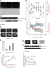
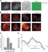
Figure 1.
Figure 1.
Invadopodium precursors form in response to EGF and mature to degrade matrix. (A and B) MTLn3 cells expressing GFP-actin were cultured on Alexa 568-FN/gelatin thick matrix and analyzed by time-lapse microscopy. (A) Formation of the precursor before degradation is shown (arrowhead). Bars, 10 μm. (B) Quantification of GFP-actin fluorescence intensity at all stages of invadopodia (blue) vs. that of underlying matrix (red). n = 3 invadopodia; three independent experiments. (C and D) Live cell time-lapse experiments using MTLn3 cells expressing TagRFP-cortactin plated on thin 488-gelatin matrix. (C) Representative montage showing the formation of an invadopodium precursor (top) precedes gelatin matrix degradation (bottom). 3 min/frame. Bar, 1 μm. (D) Quantification of the change in cortactin fluorescence intensity at invadopodia vs. that of the underlying matrix in response to EGF. n = 25 invadopodium precursors, two independent experiments. For cortactin, P < 0.05 for all times compared with 0 min. For 488-gelatin, P < 0.05 for all times compared with 0 min, except 1 min where P > 0.05. (E) Representative images of cortactin and Tks5 antibody staining of MTLn3 cells stimulated with EGF. Arrowheads indicate invadopodium precursors. Bar,10 μm. (F) Quantification of the number of invadopodium precursors per cell that contain Tks5 and cortactin after EGF stimulation. n = 40 (0 min), 37 (1 min), and 17 (3 min) cells; three independent experiments. P values are compared with 0 min. (G) Image showing that Tks5/cortactin punctate structures colocalize with matrix degradation (arrowheads). (H) Representative montage (background subtracted) showing that TagRFP-cortactin (top) and GFP-MT1-MMP (bottom) colocalize at invadopodium precursors formed in response to EGF. 20 seconds/frame. Bar, 1 μm. Quantification of (I) fluorescence intensity and (J) rate of fluorescence intensity increase of TagRFP-cortactin or GFP-MT1-MMP at invadopodium precursors formed in response to EGF. n = 33 invadopodium precursors; three independent experiments. For I, P < 0.05 for all times compared with 0 min.
Figure 2.
Figure 2.
Multiple domains of cortactin are important for matrix degradation by invadopodia. (A) Western blot of whole-cell lysates from MTLn3 cells transfected with ctrl or cortactin siRNA blotted for cortactin and β-actin. (B) Representative image of cells treated with ctrl or cortactin siRNA and stained for cortactin and F-actin to visualize invadopodium precursors (arrowheads). Bar, 10 μm. (C) Quantification of the percentage of cells that form invadopodium precursors and (D) the degradation area in cells treated with ctrl or cortactin siRNA. (E) Quantification of the number of invadopodium precursors per cell and (F) percentage of cells that form invadopodium precursors in cortactin mutant stable cell lines. P values are compared with WT and graphs show combined data from three independent experiments. (G) Quantification of the Alexa 568-FN degradation area/field normalized to WT cortactin. P values are compared with WT and n = number of fields scored: >80 from more than three independent experiments.
Figure 3.
Figure 3.
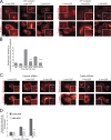
Barbed ends at invadopodium precursors peak beginning 1 min after EGF stimulation. (A) Image showing that barbed ends localize with Tks5 at areas of matrix degradation (arrowheads). Bar, 10 μm. (B and C) The barbed end assay in response to EGF using MTLn3 cells. (B) Representative images of barbed ends and F-actin at invadopodium precursors in response to EGF. Insets here and throughout the figures show close ups of invadopodium precursors. Bar, 10 μm. (C) Quantification of barbed end intensity at invadopodium precursors normalized to 0 s (sec) EGF. n = number of invadopodium precursors; three independent experiments: 0 s (39), 60 (73), 120 (109), 180 (28). P values are compared with 0 s. (D) Quantification of barbed end intensity at invadopodium precursors in response to EGF measured using a GFP-actin live cell method (Lorenz et al., 2004a). n = 21 invadopodium precursors.
Figure 4.
Figure 4.
Cortactin tyrosine phosphorylation and cofilin are important for barbed end formation at invadopodium precursors. (A and B) Barbed end assay in response to EGF with cortactin WT or 3YF cells, treated with cortactin siRNA. (A) Representative images and (B) quantification of barbed end intensity in response to EGF at invadopodium precursors normalized to WT at 0 s. Bar, 10 μm. P values are compared with WT at the same time point. Throughout the figure, n = number of invadopodium precursors; three independent experiments: WT 0 (39), 3YF 0 (24), WT 60 (255), 3YF 60 (103), WT 120 (192), 3YF 120 (171). (C) Representative images and (D) quantification of barbed ends in response to EGF in ctrl vs. cofilin siRNA treated cells normalized to 0 s. Bar, 10 μm. P values are compared with ctrl siRNA at the same time point. n: ctrl siRNA 0 (33), 60 (42), 120 (60) and cofilin siRNA 0 (57), 60 (49), 120 (110).
Figure 5.
Figure 5.
Cortactin interacts with cofilin at invadopodium precursors through a phosphorylation-dependent mechanism. (A) Representative cofilin-cortactin FRET efficiency image of a resting cell (0 s EGF). Red = cortactin, green = cofilin. Box indicates bleached area. Bottom right inset shows close up of FRET efficiency between cofilin and cortactin at invadopodium precursors. Bar, 10 μm. (B) Quantification of FRET between cofilin and cortactin at invadopodium precursors in response to EGF normalized to 0 s (FRET efficiency at 0 s = 11.8% ± 1.1). P values are compared with 0 s unless indicated. n = number of invadopodium precursors: 0 (25), 60 (24), 180 (22), 300 (17); three independent experiments. (C) Quantification of cofilin-cortactin FRET at invadopodium precursors in starved MTLn3 cells lines expressing WT, 3YF, and 3YE cortactin with endogenous cortactin knocked down normalized to WT (FRET efficiency of WT cortactin = 4.2% ± 0.36). P values are compared with WT. n = number of invadopodium precursors: WT (22), 3YF (22) 3YE (17); three independent experiments. (D–G) IP experiments between cofilin and cortactin in response to EGF. (D) Representative Western blot showing IP of cofilin and coIP of cortactin. (E) Quantification of the protein level of Co-IP cortactin/IP cofilin normalized to 0 s. n = number of independent experiments: 6. P values are compared with 0 s (F) Representative Western blot showing IP of cortactin and coIP of cofilin. (G) Quantification of the protein level of the Co-IP cofilin/IP cortactin normalized to 0 s. P values are compared with 0 s. n = number of independent experiments: 4. (H) Representative Western blot showing IP of cofilin and coIP of cortactin in cortactin WT vs. 3YF cell lines treated with cortactin siRNA. (I) Quantification of the protein level of Co-IP cortactin/IP cofilin normalized to WT. n = number of independent experiments: 3.
Figure 6.
Figure 6.
Cortactin directly binds to cofilin and inhibits cofilin's severing activity, and this inhibition is relieved when cortactin is tyrosine phosphorylated. (A and B) (left) Coomassie-stained gels and (right) quantification of the binding signal of cofilin to (A) WT cortactin (Kd = 3.5 ± 1.1 μM) or (B) 3YF cortactin (Kd = 2.8 ± 0.34 μM) from in vitro pull-down assays at increasing concentrations of cofilin. Number of data points for Kd calculation: WT = 12, 3YF = 16. (C) Representative images showing cofilin's severing activity with cofilin alone, in the presence of cortactin, or phospho-cortactin. Arrowheads show actin filaments severed by cofilin. Bar, 2 μm. (D) Quantification of the percent increase in the number of actin filaments after incubation with cofilin alone, cofilin with cortactin, or cofilin with phospho-cortactin. P > 0.05 for cofilin alone vs. cofilin with p-cortactin. (E) Quantification of the percent increase in the number of actin filaments after preincubation of actin filaments with cortactin or buffer alone as described in Materials and methods. (F) Representative cofilin/β-actin FRET efficiency images of cells stimulated with EGF for 0 (top) and 1 min (bottom). Red = β-actin, green = cofilin. Bar, 10 μm. (G) Quantification of FRET between cofilin and β-actin at invadopodium precursors in response to EGF normalized to time 0 (FRET efficiency at 0 s = 4.2% ± 0.6). n = number of invadopodium precursors: 0 (34), 60 (46); three independent experiments.
Figure 7.
Figure 7.
The tyrosine phosphorylation of cortactin is important for Nck1 localization and N-WASp activity at invadopodium precursors for Arp2/3-dependent barbed end formation. (A) Representative images of EGF-induced barbed ends at invadopodium precursors in ctrl vs. Nck1 vs. p34 KD cells. Bar, 10 μm. (B) Quantification (normalized to 0 min) of barbed ends formed in response to EGF at invadopodium precursors in ctrl vs. Nck1 vs. p34 KD cells. P values are compared with ctrl siRNA at the same time point. n = number of invadopodium precursors from two independent experiments: ctrl 0 (264), 1 (287), 2 (275), Nck1 0 (223), 1 (356), 2 (375), p34 0 (130), 1 (201), 2 (259). (C) Representative images of GFP-Nck1 and cortactin in WT and 3YF cortactin cell lines (arrowheads show invadopodium precursors). Bar, 10 μm. (D) Quantification of fold enrichment of GFP-Nck1 at invadopodium precursors in WT and 3YF cell lines. n = number of invadopodium precursors: WT (29), 3YF (34). (E) Representative Western blot showing the coIP of endogenous cortactin and pY421-cortactin with endogenous Nck1. n = 3 independent experiments. (F) Representative images of N-WASp activity measured using the CFP/YFP FRET ratio in live cells expressing cortactin WT or 3YF (arrowheads show invadopodium precursors). Bar, 10 μm. (G) Quantification of the fold enrichment of N-WASp activity at invadopodium precursors in WT and 3YF cell lines. n = number of invadopodium precursors: WT (20), 3YF (25).
Figure 8.
Figure 8.
The tyrosine dephosphorylation of cortactin is required for stable long-lived invadopodia. (A) Quantification of the average lifetimes of invadopodium precursors in cortactin WT, 3YF, and 3YE GFP-actin cell lines. n = number of invadopodium precursors: WT (133), 3YF (183), 3YE (152) from more than three independent experiments. (B) The fluorescence intensity of Arp2, cofilin, and N-WASp at invadopodium precursors in WT, 3YF, and 3YE cell lines. P values for A and B are compared with WT. n = number of invadopodium precursors: >30, three independent experiments. (C) Pearson's correlation analysis between cortactin and cofilin (r = 0.80) vs. pY421-cortactin/total cortactin and cofilin (r = −0.51) in invadopodium precursors. Error bars represent ± 95% confidence interval. (D) Quantification of the fluorescence intensity of pY421-cortactin/total cortactin at invadopodium precursors in ctrl vs. v-Src expressing cells. (E) Quantification of average invadopodium precursors lifetimes in GFP-actin cells expressing v-Src. (F) Quantification of the average FN degradation area/field, (G) number of invadopodium precursors/cell, and (H) FN degradation area normalized to the number of invadopodium precursors/cell in ctrl vs. v-Src expressing cells.
Figure 9.
Figure 9.
Cortactin regulates the activities of cofilin and N-WASp to control the stages of invadopodium assembly and maturation (model). During precursor formation (stage 1), cortactin, N-WASp, cofilin, and Arp2/3 form a complex involving cortactin's Arp2/3 and N-WASp binding domains. Cortactin is then tyrosine phosphorylated, which activates cofilin's severing activity to generate free barbed ends and the Arp2/3 complex can use these cofilin-generated barbed ends for efficient actin polymerization (stage 2). Cortactin is then dephosphorylated, which stabilizes the invadopodium precursor for maturation (stage 3). Box at stage 3 and 4 indicates stabilization. MT1-MMP can be recruited at stages 2–4. Stages 1–3 are required for a precursor to become a mature invadopodium that efficienctly degrades ECM (stage 4).

References

    1. Artym V.V., Zhang Y., Seillier-Moiseiwitsch F., Yamada K.M., Mueller S.C. 2006. Dynamic interactions of cortactin and membrane type 1 matrix metalloproteinase at invadopodia: defining the stages of invadopodia formation and function.Cancer Res. 66:3034–3043 - PubMed
    1. Ayala I., Baldassarre M., Giacchetti G., Caldieri G., Tetè S., Luini A., Buccione R. 2008. Multiple regulatory inputs converge on cortactin to control invadopodia biogenesis and extracellular matrix degradation.J. Cell Sci. 121:369–378 - PubMed (VSports最新版本)
    1. Boyle S.N., Michaud G.A., Schweitzer B., Predki P.F., Koleske A.J. 2007. A critical role for cortactin phosphorylation by Abl-family kinases in PDGF-induced dorsal-wave formation.Curr. Biol. 17:445–451 - PubMed (V体育安卓版)
    1. Bravo-Cordero J.J., Marrero-Diaz R., Megías D., Genís L., García-Grande A., García M.A., Arroyo A.G., Montoya M.C. 2007. MT1-MMP proinvasive activity is regulated by a novel Rab8-dependent exocytic pathway.EMBO J. 26:1499–1510 - PMC - PubMed
    1. Bryce N.S., Clark E.S., Leysath J.L., Currie J.D., Webb D.J., Weaver A.M. 2005. Cortactin promotes cell motility by enhancing lamellipodial persistence.Curr. Biol. 15:1276–1285 - PubMed

Publication types

MeSH terms (VSports最新版本)

Substances